

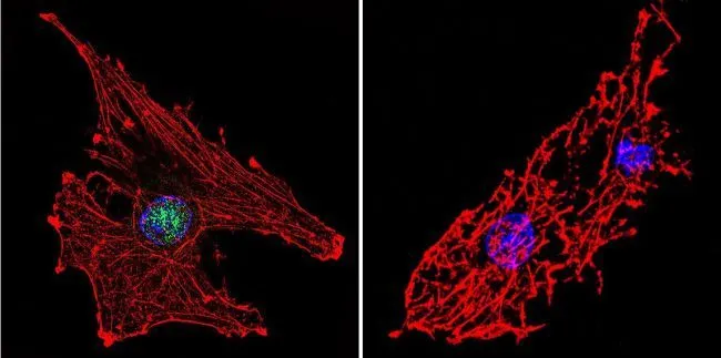
ICC/IF analysis of C6 cells using GTX15783 RPA32 antibody [MA34]. Cells were probed without (right) or with(left) an antibody. Green : Primary antibody Blue : Nuclei Red : Actin Fixation : formaldehyde Dilution : 1:200 overnight at 4oC
![IHC-P analysis of human tonsil tissue using GTX15783 RPA32 antibody [MA34]. Left : Primary antibody Right : Negative control without primary antibody Antigen retrieval : heat induced antigen retrieval was performed using 10mM sodium citrate (pH6.0) buffer, microwaved for 8-15 minutes Dilution : 1:20](https://www.genetex.com/upload/website/prouct_img/normal/GTX15783/GTX15783_1043_IHC-P_w_23060620_158.webp "IHC-P analysis of human tonsil tissue using GTX15783 RPA32 antibody [MA34]. Left : Primary antibody Right : Negative control without primary antibody Antigen retrieval : heat induced antigen retrieval was performed using 10mM sodium citrate (pH6.0) buffer, microwaved for 8-15 minutes Dilution : 1:20")
![IHC-P analysis of human kidney tissue using GTX15783 RPA32 antibody [MA34]. Left : Primary antibody Right : Negative control without primary antibody Antigen retrieval : heat induced antigen retrieval was performed using 10mM sodium citrate (pH6.0) buffer, microwaved for 8-15 minutes Dilution : 1:20](https://www.genetex.com/upload/website/prouct_img/normal/GTX15783/GTX15783_1042_IHC-P_w_23060620_365.webp "IHC-P analysis of human kidney tissue using GTX15783 RPA32 antibody [MA34]. Left : Primary antibody Right : Negative control without primary antibody Antigen retrieval : heat induced antigen retrieval was performed using 10mM sodium citrate (pH6.0) buffer, microwaved for 8-15 minutes Dilution : 1:20")
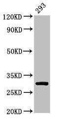
![WB analysis of purified RPA protein using GTX15783 RPA32 antibody [MA34].](https://www.genetex.com/upload/website/prouct_img/normal/GTX15783/GTX15783_1535_WB_w_23060620_654.webp "WB analysis of purified RPA protein using GTX15783 RPA32 antibody [MA34].")
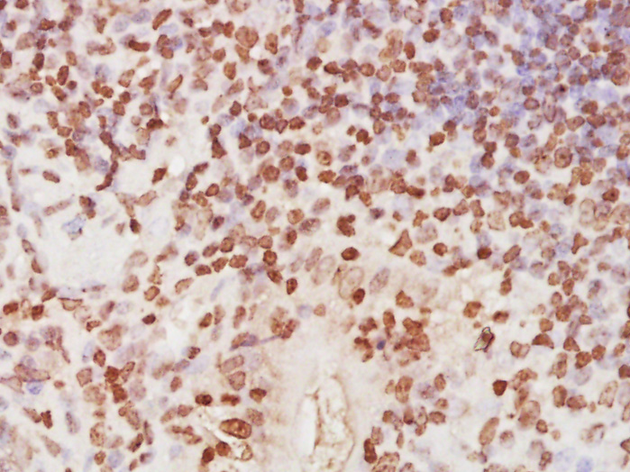
![IHC-P analysis of human breast carcinoma tissue using GTX15783 RPA32 antibody [MA34]. Left : Primary antibody Right : Negative control without primary antibody Antigen retrieval : heat induced antigen retrieval was performed using 10mM sodium citrate (pH6.0) buffer, microwaved for 8-15 minutes Dilution : 1:20](https://www.genetex.com/upload/website/prouct_img/normal/GTX15783/GTX15783_1041_IHC-P_w_23060620_950.webp "IHC-P analysis of human breast carcinoma tissue using GTX15783 RPA32 antibody [MA34]. Left : Primary antibody Right : Negative control without primary antibody Antigen retrieval : heat induced antigen retrieval was performed using 10mM sodium citrate (pH6.0) buffer, microwaved for 8-15 minutes Dilution : 1:20")
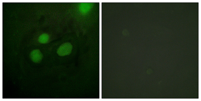
![ICC/IF analysis of A431 cells using GTX15783 RPA32 antibody [MA34]. Cells were probed without (right) or with(left) an antibody. Green : Primary antibody Blue : Nuclei Red : Actin Fixation : formaldehyde Dilution : 1:200 overnight at 4oC](https://www.genetex.com/upload/website/prouct_img/normal/GTX15783/GTX15783_313_ICC-IF_w_23060620_419.webp "ICC/IF analysis of A431 cells using GTX15783 RPA32 antibody [MA34]. Cells were probed without (right) or with(left) an antibody. Green : Primary antibody Blue : Nuclei Red : Actin Fixation : formaldehyde Dilution : 1:200 overnight at 4oC")
![ICC/IF analysis of HeLa cells using GTX15783 RPA32 antibody [MA34]. Cells were probed without (right) or with(left) an antibody. Green : Primary antibody Blue : Nuclei Red : Actin Fixation : formaldehyde Dilution : 1:200 overnight at 4oC](https://www.genetex.com/upload/website/prouct_img/normal/GTX15783/GTX15783_315_ICC-IF_w_23060620_646.webp "ICC/IF analysis of HeLa cells using GTX15783 RPA32 antibody [MA34]. Cells were probed without (right) or with(left) an antibody. Green : Primary antibody Blue : Nuclei Red : Actin Fixation : formaldehyde Dilution : 1:200 overnight at 4oC")
ICC/IF analysis of C6 cells using GTX15783 RPA32 antibody [MA34]. Cells were probed without (right) or with(left) an antibody. Green : Primary antibody Blue : Nuclei Red : Actin Fixation : formaldehyde Dilution : 1:200 overnight at 4oC
RPA32 antibody [MA34]
GTX15783
ApplicationsImmunoFluorescence, Western Blot, ImmunoCytoChemistry, ImmunoHistoChemistry, ImmunoHistoChemistry Paraffin
Product group Antibodies
ReactivityHuman, Rat
TargetRPA2
Overview
- SupplierGeneTex
- Product NameRPA32 antibody [MA34]
- Delivery Days Customer9
- Application Supplier NoteWB: 1:500. ICC/IF: 1:100-1:1000. IHC-P: 1:10-1:100. *Optimal dilutions/concentrations should be determined by the researcher.Not tested in other applications.
- ApplicationsImmunoFluorescence, Western Blot, ImmunoCytoChemistry, ImmunoHistoChemistry, ImmunoHistoChemistry Paraffin
- CertificationResearch Use Only
- ClonalityMonoclonal
- Clone IDMA34
- Concentration1 mg/ml
- ConjugateUnconjugated
- Gene ID6118
- Target nameRPA2
- Target descriptionreplication protein A2
- Target synonymsREPA2, RP-A p32, RP-A p34, RPA32, replication protein A 32 kDa subunit, RF-A protein 2, replication factor A protein 2, replication protein A 34 kDa subunit
- HostMouse
- IsotypeIgM
- Protein IDP15927
- Protein NameReplication protein A 32 kDa subunit
- Scientific DescriptionThis gene encodes a subunit of the heterotrimeric Replication Protein A (RPA) complex, which binds to single-stranded DNA (ssDNA), forming a nucleoprotein complex that plays an important role in DNA metabolism, being involved in DNA replication, repair, recombination, telomere maintenance, and co-ordinating the cellular response to DNA damage through activation of the ataxia telangiectasia and Rad3-related protein (ATR) kinase. The RPA complex protects single-stranded DNA from nucleases, prevents formation of secondary structures that would interfere with repair, and co-ordinates the recruitment and departure of different genome maintenance factors. The heterotrimeric complex has two different modes of ssDNA binding, a low-affinity and high-affinity mode, determined by which oligonucleotide/oligosaccharide-binding (OB) domains of the complex are utilized, and differing in the length of DNA bound. This subunit contains a single OB domain that participates in high-affinity DNA binding and also contains a winged helix domain at its carboxy terminus, which interacts with many genome maintenance protein. Post-translational modifications of the RPA complex also plays a role in co-ordinating different damage response pathways. [provided by RefSeq, Sep 2017]
- ReactivityHuman, Rat
- Storage Instruction-20°C or -80°C,2°C to 8°C
- UNSPSC41116161
Datasheet
Related products
Product group Antibodies
Anti-RFA2 AntibodyA96134
ApplicationsImmunoFluorescence, Western Blot, ELISA, ImmunoHistoChemistry
ReactivityHuman, Mouse
- SizePrice
Product group Antibodies
Anti-RFA2 Antibody102-20182
ApplicationsWestern Blot, ImmunoHistoChemistry, ImmunoHistoChemistry Paraffin
TargetRPA2
- SizePrice
Product group Antibodies

RPA2 / RFA2 / RPA34 AntibodyLS-C770317
ApplicationsWestern Blot, ELISA, ImmunoHistoChemistry
ReactivityHuman
TargetRPA2
- SizePrice

Product group Antibodies
Anti-RPA32/RPA2 Antibody Picoband(r)A02067-1-CARRIER-FREE
ApplicationsFlow Cytometry, ImmunoFluorescence, Western Blot, ELISA, ImmunoCytoChemistry, ImmunoHistoChemistry
ReactivityHuman
TargetRPA2
- SizePrice
Product group Antibodies
RPA2 Polyclonal AntibodyBS-4182R
ApplicationsImmunoFluorescence, Western Blot, ELISA, ImmunoCytoChemistry, ImmunoHistoChemistry, ImmunoHistoChemistry Frozen, ImmunoHistoChemistry Paraffin
ReactivityBovine, Canine, Chicken, Equine, Human, Mouse, Porcine, Rabbit, Rat
TargetRPA2
- SizePrice
Product group Antibodies
RPA2 Polyclonal AntibodyCAC13840
ApplicationsImmunoFluorescence, ImmunoPrecipitation, Western Blot, ELISA, ImmunoHistoChemistry
TargetRPA2
- SizePrice
Product group Antibodies
RPA2 AntibodyCSB-PA01555A0RB
ApplicationsImmunoFluorescence, ImmunoPrecipitation, Western Blot, ELISA, ImmunoHistoChemistry
ReactivityHuman
TargetRPA2
- SizePrice
Product group Antibodies
References
RPA32 antibody [RPA34-19]GTX16855
ApplicationsImmunoFluorescence, ImmunoPrecipitation, Western Blot, ImmunoCytoChemistry, ImmunoHistoChemistry, ImmunoHistoChemistry Paraffin
ReactivityHamster, Human
TargetRPA2
- SizePrice
![IHC-P analysis of human tonsil tissue using GTX22175 RPA32 antibody [9H8].](https://www.genetex.com/upload/website/prouct_img/normal/GTX22175/GTX22175_20191203_IHC-P_94_w_23060620_908.webp)
Product group Antibodies
References
RPA32 antibody [9H8]GTX22175
ApplicationsImmunoFluorescence, Western Blot, ImmunoCytoChemistry, ImmunoHistoChemistry, ImmunoHistoChemistry Paraffin
ReactivityHuman, Monkey
TargetRPA2
- SizePrice
![WB analysis of various samples using GTX02702 RPA32 antibody [RPA2/3140R]. Lane 1 : MCF-7 whole cell lysate Lane 2 : T47D cell lysate](https://www.genetex.com/upload/website/prouct_img/normal/GTX02702/GTX02702_20210319_WB_w_23053122_247.webp)
Product group Antibodies
RPA32 antibody [RPA2/3140R]GTX02702
ApplicationsWestern Blot
ReactivityHuman
TargetRPA2
- SizePrice