

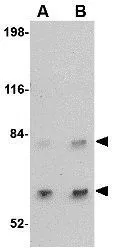
WB analysis of A-20 cell lysate using GTX30894 SATB2 antibody. Working concentration : (A) 2 and (B) 4 μg/ml


WB analysis of A-20 cell lysate using GTX30894 SATB2 antibody. Working concentration : (A) 2 and (B) 4 μg/ml
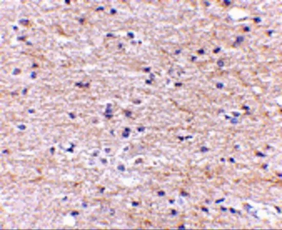
SATB2 antibody
GTX30894
ApplicationsWestern Blot, ELISA, ImmunoHistoChemistry, ImmunoHistoChemistry Paraffin
Product group Antibodies
ReactivityHuman, Mouse, Rat
TargetSATB2
Overview
- SupplierGeneTex
- Product NameSATB2 antibody
- Delivery Days Customer9
- Application Supplier NoteWB: 2 - 4 microg/mL. IHC-P: 5 microg/mL. *Optimal dilutions/concentrations should be determined by the researcher.Not tested in other applications.
- ApplicationsWestern Blot, ELISA, ImmunoHistoChemistry, ImmunoHistoChemistry Paraffin
- CertificationResearch Use Only
- ClonalityPolyclonal
- Concentration1 mg/ml
- ConjugateUnconjugated
- Gene ID23314
- Target nameSATB2
- Target descriptionSATB homeobox 2
- Target synonymsC2DELq32q33, DEL2Q32Q33, GLSS, DNA-binding protein SATB2, SATB family member 2, SATB2 fusion, special AT-rich sequence-binding protein 2
- HostRabbit
- IsotypeIgG
- Protein IDQ9UPW6
- Protein NameDNA-binding protein SATB2
- Scientific DescriptionHuman special AT-rich sequence-binding protein-2 (SATB2) is a nuclear matrix/scaffold-associated region DNA-binding protein. Like its homolog SATB1, SATB2 selectively binds double-stranded, special AT-rich DNA sequences, but is expressed primarily in a subset of postmitotic, differentiating neurons in the neocortex. Mice deficient in SATB exhibit craniofacial abnormalities and defects in osteoblast differentiation and function. SATB2 also interacts with and enhances the activity of Runx2 and ATF4, two transcription factors that regulate osteoblast differentiation, indicating that SATB2 acts as a molecular node in a transcriptional network regulating skeletal development and osteoblast differentiation. Recent experiments have shown that SATB2 interacts with histone deacetylase 1 and metastasis-associated protein 2, two proteins that are involved in chromatin remodeling, suggesting that SATB2 may also be involved in mediating epigenetic influences during cortical development. At least two isoforms of SATB2 are known to exist.
- ReactivityHuman, Mouse, Rat
- Storage Instruction-20°C or -80°C,2°C to 8°C
- UNSPSC41116161
Datasheet
Related products
Product group Antibodies
Anti-SATB2 AntibodyA91312
ApplicationsWestern Blot, ImmunoHistoChemistry
ReactivityHuman, Rat
- SizePrice

Product group Antibodies
Anti-SATB2 Antibody Picoband(r)A02588-2-CARRIER-FREE
ApplicationsWestern Blot
ReactivityHuman
TargetSATB2
- SizePrice
Product group Antibodies
Anti-SATB2 Antibody144-64612
ApplicationsWestern Blot
ReactivityHuman
TargetSATB2
- SizePrice

Product group Antibodies
Anti-SATB2 AntibodyAMAB90678
ApplicationsWestern Blot, ImmunoHistoChemistry
ReactivityHuman, Rat
TargetSATB2
- SizePrice
Product group Antibodies
References
SATB2 Polyclonal AntibodyBS-11949R
ApplicationsFlow Cytometry, ImmunoFluorescence, Western Blot, ELISA, ImmunoCytoChemistry, ImmunoHistoChemistry, ImmunoHistoChemistry Frozen, ImmunoHistoChemistry Paraffin
ReactivityBovine, Equine, Human, Mouse, Rat, Sheep
TargetSATB2
- SizePrice
Product group Antibodies
SATB2 AntibodyCSB-PA892170LA01HU
ApplicationsELISA, ImmunoHistoChemistry
ReactivityHuman
TargetSATB2
- SizePrice
Product group Antibodies
ApplicationsImmunoPrecipitation, Western Blot, ImmunoCytoChemistry, ImmunoHistoChemistry
TargetSATB2
- SizePrice

Product group Antibodies
SATB2 antibodyGTX132972
ApplicationsImmunoFluorescence, Western Blot, ImmunoCytoChemistry
ReactivityHuman, Rat
TargetSATB2
- SizePrice
Product group Antibodies
SATB2 antibodyGTX31773
ApplicationsWestern Blot, ELISA, ImmunoHistoChemistry, ImmunoHistoChemistry Paraffin
ReactivityHuman, Mouse, Rat
TargetSATB2
- SizePrice