

Search results: akt
Product group Antibodies
ApplicationsFlow Cytometry, ImmunoFluorescence, Western Blot, ELISA, ImmunoHistoChemistry
ReactivityHuman, Mouse
TargetAKT1
- SizePrice
Product group Antibodies
ApplicationsFlow Cytometry, Western Blot, ELISA, ImmunoHistoChemistry
ReactivityHuman, Mouse
TargetAKT1
- SizePrice
Product group Antibodies
ApplicationsFlow Cytometry, Western Blot, ELISA, ImmunoHistoChemistry
ReactivityMouse
TargetAKT1
- SizePrice
Product group Antibodies
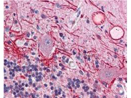
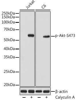
ApplicationsWestern Blot, ImmunoHistoChemistry
ReactivityHuman, Mouse, Rat
TargetAKT1
- SizePrice
Product group Antibodies
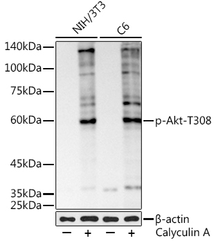
ApplicationsWestern Blot, ImmunoHistoChemistry
ReactivityHuman
TargetAKT1
- SizePrice
Product group Antibodies
AKT1 Rabbit Polyclonal AntibodyTA420004
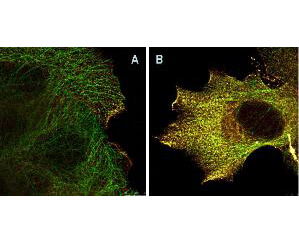
ApplicationsImmunoFluorescence, ImmunoPrecipitation, Western Blot, ImmunoCytoChemistry, ImmunoHistoChemistry
ReactivityHuman, Mouse, Rat
TargetAKT1
- SizePrice
Product group Antibodies
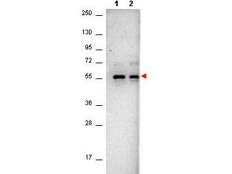
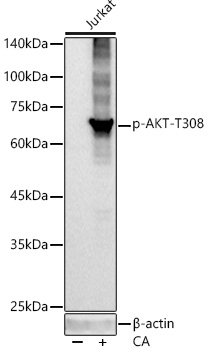
ApplicationsWestern Blot
ReactivityHuman, Mouse, Rat
TargetAKT1
- SizePrice
Product group Antibodies
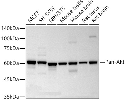
ApplicationsWestern Blot
ReactivityMouse, Rat
TargetAKT1
- SizePrice
Product group Antibodies
ApplicationsWestern Blot, ImmunoHistoChemistry
ReactivityHuman, Mouse, Rat
TargetAKT1
- SizePrice
![17-AAG [75747-14-7]](https://bpsbioscience.com/media/catalog/product/2/7/27027.jpg)
Product group Chemicals
17-AAG [75747-14-7]27027
CAS Number75747-14-7
Estimated Purity≥99%
Molecular Weight585.7 Da
- SizePrice
Product group Chemicals
PP242 [1092351-67-1]27063
CAS Number1092351-67-1
Estimated Purity≥99%
Molecular Weight308.3 Da
- SizePrice
![Chidamide [743420-02-2]](https://bpsbioscience.com/media/catalog/product/2/7/27202_structure.png)
Product group Chemicals
CAS Number743420-02-2
Estimated Purity≥98%
Molecular Weight390.4 Da
- SizePrice
Didn't find what you were looking for?
Search through our product groups to find the right product
Back to overview