

SEMA7A antibody [MEM-150] (FITC)
GTX28222-06
ApplicationsFlow Cytometry
Product group Antibodies
ReactivityHuman
TargetSEMA7A
Overview
- SupplierGeneTex
- Product NameSEMA7A antibody [MEM-150] (FITC)
- Delivery Days Customer9
- Application Supplier NoteFCM: 20 microl reagent / 100 microl of whole blood or 106 cells in a suspension. *Optimal dilutions/concentrations should be determined by the researcher.Not tested in other applications.
- ApplicationsFlow Cytometry
- CertificationResearch Use Only
- ClonalityMonoclonal
- Clone IDMEM-150
- ConjugateFITC
- Gene ID8482
- Target nameSEMA7A
- Target descriptionsemaphorin 7A (JohnMiltonHagen blood group)
- Target synonymsCD108, CDw108, H-SEMA-K1, H-Sema-L, JMH, PFIC11, SEMAK1, SEMAL, semaphorin-7A, JMH blood group antigen, John Milton Hagen blood group H-Sema K1, john-Milton-Hargen human blood group Ag, sema K1, sema L, sema domain, immunoglobulin domain (Ig), and GPI membrane anchor, (semaphorin) 7A (JMH blood group), sema domain, immunoglobulin domain (Ig), and GPI membrane anchor, 7A, semaphorin 7A (John Milton Hagen blood group), semaphorin 7A, GPI membrane anchor (John Milton Hagen blood group), semaphorin-K1, semaphorin-L
- HostMouse
- IsotypeIgM
- Protein IDO75326
- Protein NameSemaphorin-7A
- Scientific DescriptionThis gene encodes a member of the semaphorin family of proteins. The encoded preproprotein is proteolytically processed to generate the mature glycosylphosphatidylinositol (GPI)-anchored membrane glycoprotein. The encoded protein is found on activated lymphocytes and erythrocytes and may be involved in immunomodulatory and neuronal processes. The encoded protein carries the John Milton Hagen (JMH) blood group antigens. Mutations in this gene may be associated with reduced bone mineral density (BMD). Alternative splicing results in multiple transcript variants, at least one of which encodes an isoform that is proteolytically processed. [provided by RefSeq, Feb 2016]
- ReactivityHuman
- Storage Instruction2°C to 8°C
- UNSPSC41116161
Datasheet
Related products
Product group Antibodies
Anti-SEMA7A AntibodyA37320
ApplicationsWestern Blot, ImmunoHistoChemistry
ReactivityHuman, Mouse
- SizePrice

Product group Antibodies
Anti-Semaphorin 7a/SEMA7A Antibody Picoband(r)A03832-2-CARRIER-FREE
ApplicationsFlow Cytometry, Western Blot, ELISA
ReactivityHuman
TargetSEMA7A
- SizePrice
Product group Antibodies
Anti-SEMA7A Antibody144-62182
ApplicationsWestern Blot
ReactivityHuman
TargetSEMA7A
- SizePrice
Product group Antibodies

SEMA7A / Semaphorin 7A AntibodyLS-C747248
ApplicationsWestern Blot
ReactivityHuman
TargetSEMA7A
- SizePrice
Product group Antibodies
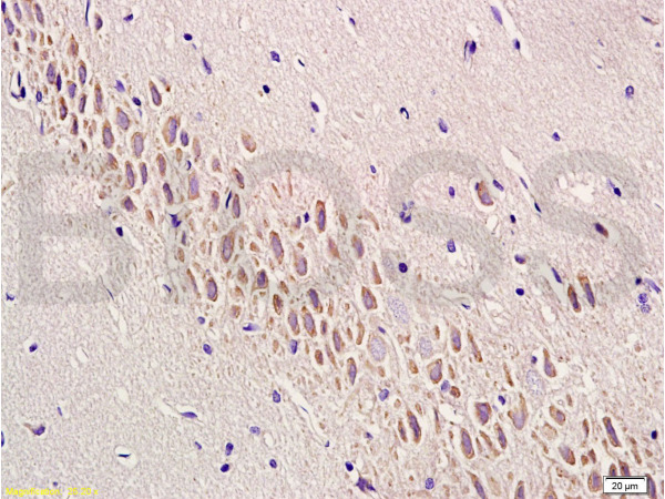
CD108 Polyclonal AntibodyBS-2702R
ApplicationsImmunoFluorescence, Western Blot, ELISA, ImmunoCytoChemistry, ImmunoHistoChemistry, ImmunoHistoChemistry Frozen, ImmunoHistoChemistry Paraffin
ReactivityBovine, Canine, Human, Mouse, Rat
TargetSEMA7A
- SizePrice
Product group Antibodies
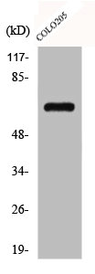
SEMA7A AntibodyCSB-PA001418
ApplicationsWestern Blot, ELISA
ReactivityHuman, Mouse, Rat
TargetSEMA7A
- SizePrice
Product group Antibodies
ApplicationsImmunoPrecipitation, Western Blot, ImmunoCytoChemistry, ImmunoHistoChemistry
ReactivityPorcine
TargetSEMA7A
- SizePrice
![FACS analysis of human peripheral blood using GTX28222 SEMA7A antibody [MEM-150].](https://www.genetex.com/upload/website/prouct_img/normal/GTX28222/GTX28222_20191028_WB_1_w_23060722_221.webp)
Product group Antibodies
SEMA7A antibody [MEM-150]GTX28222
ApplicationsFlow Cytometry, ImmunoPrecipitation, Western Blot
ReactivityHuman
TargetSEMA7A
- SizePrice

Product group Antibodies
Anti-SEMA7A AntibodyHPA042273
ApplicationsImmunoHistoChemistry
ReactivityHuman
TargetSEMA7A
- SizePrice
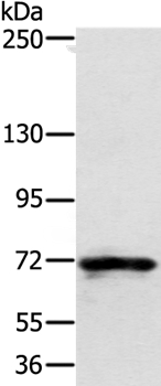
![WB analysis of human brain tissue lysate using GTX52991 SEMA7A antibody [99D22].](https://www.genetex.com/upload/website/prouct_img/normal/GTX52991/GTX52991_20191119_WB_w_23060900_591.webp)
Product group Antibodies
SEMA7A antibody [99D22]GTX52991
ApplicationsWestern Blot
ReactivityHuman
TargetSEMA7A
- SizePrice