

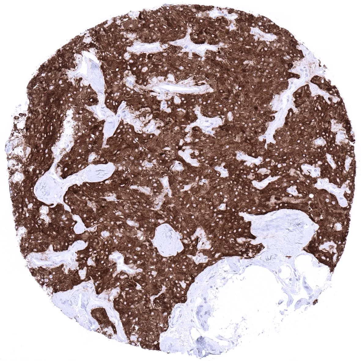
IHC-P analysis of human medullary thyroid cancer (MTC) tissue using GTX04371 Synaptophysin antibody [MSVA-462R] HistoMAX?. Strong cytoplasmic synaptophysin immunostaining of all tumor cells.
![IHC-P analysis of human pancreas tissue using GTX04371 Synaptophysin antibody [MSVA-462R] HistoMAX?. A strong synaptophysin immunostaining is seen in islet cells of Langerhans.](https://www.genetex.com/upload/website/prouct_img/normal/GTX04371/GTX04371_20230728_IHC-P_235_23072723_306.webp "IHC-P analysis of human pancreas tissue using GTX04371 Synaptophysin antibody [MSVA-462R] HistoMAX?. A strong synaptophysin immunostaining is seen in islet cells of Langerhans.")
![IHC-P analysis of human cerebellum (molecular layer, Purkinje cell layer, granule cell layer, and white matter) tissue using GTX04371 Synaptophysin antibody [MSVA-462R] HistoMAX?.
Synaptophysin is strongly expressed in cerebellum molecular, Purkinje and granular layer.](https://www.genetex.com/upload/website/prouct_img/normal/GTX04371/GTX04371_20230728_IHC-P_365_23072723_311.webp "IHC-P analysis of human cerebellum (molecular layer, Purkinje cell layer, granule cell layer, and white matter) tissue using GTX04371 Synaptophysin antibody [MSVA-462R] HistoMAX?.
Synaptophysin is strongly expressed in cerebellum molecular, Purkinje and granular layer.")
IHC-P analysis of human medullary thyroid cancer (MTC) tissue using GTX04371 Synaptophysin antibody [MSVA-462R] HistoMAX?. Strong cytoplasmic synaptophysin immunostaining of all tumor cells.
Synaptophysin antibody [MSVA-462R] HistoMAX(tm)
GTX04371
ApplicationsImmunoHistoChemistry, ImmunoHistoChemistry Paraffin
Product group Antibodies
ReactivityHuman
TargetSYP
Overview
- SupplierGeneTex
- Product NameSynaptophysin antibody [MSVA-462R] HistoMAX(tm)
- Delivery Days Customer9
- Application Supplier NoteIHC-P: 1:100-1:200. *Optimal dilutions/concentrations should be determined by the researcher.Not tested in other applications.
- ApplicationsImmunoHistoChemistry, ImmunoHistoChemistry Paraffin
- CertificationResearch Use Only
- ClonalityMonoclonal
- Clone IDMSVA-462R
- Concentration0.2 mg/ml
- ConjugateUnconjugated
- Gene ID6855
- Target nameSYP
- Target descriptionsynaptophysin
- Target synonymsMRX96, MRXSYP, XLID96, synaptophysin, major synaptic vesicle protein P38
- HostRabbit
- IsotypeIgG
- Protein IDP08247
- Protein NameSynaptophysin
- Scientific DescriptionThis gene encodes an integral membrane protein of small synaptic vesicles in brain and endocrine cells. The protein also binds cholesterol and is thought to direct targeting of vesicle-associated membrane protein 2 (synaptobrevin) to intracellular compartments. Mutations in this gene are associated with an X-linked form of cognitive disability. [provided by RefSeq, Jul 2017]
- ReactivityHuman
- Storage Instruction-20°C or -80°C,2°C to 8°C
- UNSPSC12352203
Datasheet
Related products
Product group Antibodies

Synaptophysin (SYP) AntibodyABX013199
ApplicationsWestern Blot, ELISA, ImmunoHistoChemistry
- SizePrice
Product group Antibodies
Anti-SYP Antibody144-06344
ApplicationsImmunoFluorescence, Western Blot, ImmunoHistoChemistry
ReactivityHuman, Mouse, Rat
TargetSYP
- SizePrice
![Synaptophysin antibody detects Synaptophysin protein at synaptic vesicles by immunofluorescent analysis. Sample: DIV9 rat E18 primary cortical neurons were fixed in 4% paraformaldehyde at RT for 15 min. Green: Synaptophysin protein stained by Synaptophysin antibody (GTX100865) diluted at 1:500. Red: beta Tubulin 3/ Tuj1, stained by beta Tubulin 3/ Tuj1 antibody [GT11710] (GTX631836) diluted at 1:500. Blue: Fluoroshield with DAPI (GTX30920).](https://www.genetex.com/upload/website/prouct_img/normal/GTX100865/GTX100865_40128_20170503_IFA_R_w_23060100_733.webp)
Product group Antibodies
References
Synaptophysin antibodyGTX100865
ApplicationsImmunoFluorescence, Western Blot, ImmunoCytoChemistry, ImmunoHistoChemistry, ImmunoHistoChemistry Frozen, ImmunoHistoChemistry Paraffin
ReactivityHuman, Mouse, Rat
TargetSYP
- SizePrice
![Synaptophysin antibody [GT1825] detects Synaptophysin protein at cell membrane in mouse brain by immunohistochemical analysis. Sample: Paraffin-embedded mouse brain. Synaptophysin antibody [GT1825] (GTX633821) diluted at 1:200.
Antigen Retrieval: Citrate buffer, pH 6.0, 15 min](https://www.genetex.com/upload/website/prouct_img/normal/GTX633821/GTX633821_42667_20170329_IHC-P_M_w_23061202_125.webp)
Product group Antibodies
References
Synaptophysin antibody [GT1825]GTX633821
ApplicationsWestern Blot, ImmunoHistoChemistry, ImmunoHistoChemistry Frozen, ImmunoHistoChemistry Paraffin
ReactivityHuman, Mouse, Rat
TargetSYP
- SizePrice
![Various tissue extracts (50 μg) were separated by 12% SDS-PAGE, and the membrane was blotted with Synaptophysin antibody [GT2589] (GTX633972) diluted at 1:10000. The HRP-conjugated anti-mouse IgG antibody (GTX213111-01) was used to detect the primary antibody.](https://www.genetex.com/upload/website/prouct_img/normal/GTX633972/GTX633972_42758_20200403_WB_M_tissue_competitor_watermark_w_23061202_715.webp)
Product group Antibodies
Synaptophysin antibody [GT2589]GTX633972
ApplicationsImmunoFluorescence, Western Blot, ImmunoCytoChemistry, ImmunoHistoChemistry, ImmunoHistoChemistry Paraffin
ReactivityHuman, Mouse, Rat
TargetSYP
- SizePrice
![IHC-P analysis of human pancreas tissue using GTX79421 Synaptophysin antibody [SP11].](https://www.genetex.com/upload/website/prouct_img/normal/GTX79421/GTX79421_20191203_IHC-P_221_w_23061322_825.webp)
Product group Antibodies
Synaptophysin antibody [SP11]GTX79421
ApplicationsImmunoHistoChemistry, ImmunoHistoChemistry Paraffin
ReactivityHuman
TargetSYP
- SizePrice
![IHC-P analysis of human pancreas tissue using GTX79435 Synaptophysin antibody [SP11] (ready-to-use).](https://www.genetex.com/upload/website/prouct_img/normal/GTX79435/GTX79435_20191203_IHC-P_230_w_23061322_607.webp)
Product group Antibodies
ApplicationsImmunoHistoChemistry, ImmunoHistoChemistry Paraffin
ReactivityHuman
TargetSYP
- SizePrice
Product group Antibodies
ApplicationsImmunoPrecipitation, Western Blot, ImmunoCytoChemistry, ImmunoHistoChemistry
TargetSYP
- SizePrice

Product group Antibodies
Synaptophysin Recombinant AntibodyBSM-52379R
ApplicationsFlow Cytometry, ImmunoFluorescence, Western Blot, ImmunoCytoChemistry, ImmunoHistoChemistry, ImmunoHistoChemistry Frozen, ImmunoHistoChemistry Paraffin
ReactivityHuman, Mouse, Rat
TargetSYP
- SizePrice