

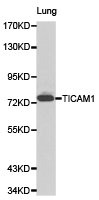
Western blot analysis of lung cell lysate using TICAM1 antibody.
Western blot analysis of lung cell lysate using TICAM1 antibody.
TICAM1 Antibody
CSB-PA023532KA01HU
ApplicationsWestern Blot, ELISA, ImmunoHistoChemistry
Product group Antibodies
ReactivityHuman, Mouse, Rat
TargetTICAM1
Overview
- SupplierCusabio
- Product NameTICAM1 Antibody
- Delivery Days Customer20
- ApplicationsWestern Blot, ELISA, ImmunoHistoChemistry
- CertificationResearch Use Only
- ClonalityPolyclonal
- ConjugateUnconjugated
- Gene ID148022
- Target nameTICAM1
- Target descriptionTIR domain containing adaptor molecule 1
- Target synonymsIIAE6, MyD88-3, PRVTIRB, TICAM-1, TRIF, TIR domain-containing adapter molecule 1, TIR domain containing adaptor inducing interferon-beta, TIR domain-containing adapter protein inducing IFN-beta, proline-rich, vinculin and TIR domain-containing protein B, putative NF-kappa-B-activating protein 502H, toll like receptor adaptor molecule 1, toll-interleukin-1 receptor domain-containing adapter protein inducing interferon beta
- HostRabbit
- IsotypeIgG
- Protein IDQ8IUC6
- Protein NameTIR domain-containing adapter molecule 1
- Scientific DescriptionMembers of the Toll-like receptor (TLR) family, named for the closely related Toll receptor in Drosophila, play a pivotal role in innate immune responses. TLRs recognize conserved motifs found in various pathogens and mediate defense responses. Triggering of the TLR pathway leads to the activation of NF-kappaB and subsequent regulation of immune and inflammatory genes. The TLRs and members of the IL-1 receptor family share a conserved stretch of approximately 200 amino acids known as the TIR domain. Upon activation, TLRs associate with a number of cytoplasmic adaptor proteins containing TIR domains including MyD88 (myeloid differentiation factor), MAL/TIRAP (MyD88-adaptor-like/TIR-associated protein), TRIF (Toll-receptor-associated activator of interferon), and TRAM (Toll-receptor-associated molecule). This association leads to the recruitment and activation of IRAK1 and IRAK4, which form a complex with TRAF6 to activate TAK1 and IKK. Activation of IKK leads to the degradation of IkappaB that normally maintains NF-kappaB inactivity by sequestering it in the cytoplasm.TRIF (also termed TICAM-1) is a TIR-domain adaptor protein described to activate NF-kappaB, IRF3 and trigger IFN-beta production. Studies using dominant negative forms of TRIF and siRNA targeting TRIF show that TRIF functions downstream of both TLR3 and TLR4 in response to dsRNA and LPS respectively. TRIF recruits TRAF6-TAK1-TAB2 to the receptor complex which leads to NF-kappaB activation. In addition, TRIF can trigger signaling of that lead to the induction of apoptosis.
- ReactivityHuman, Mouse, Rat
- Storage Instruction-20°C or -80°C
- UNSPSC41116161
Related products
Product group Antibodies
ApplicationsImmunoPrecipitation, Western Blot, ImmunoCytoChemistry, ImmunoHistoChemistry
ReactivityMouse
TargetTICAM1
- SizePrice
Product group Antibodies
TICAM1 AntibodyCSB-PA544544
ApplicationsWestern Blot, ELISA
ReactivityHuman
TargetTICAM1
- SizePrice

Product group Antibodies
Anti-TRIF/TICAM1 Antibody Picoband(r)A01872-1-CARRIER-FREE
ApplicationsFlow Cytometry, Western Blot, ELISA
ReactivityHuman
TargetTICAM1
- SizePrice
Product group Antibodies
Anti-TICAM1 Antibody144-60070
ApplicationsWestern Blot
ReactivityHuman, Mouse, Rat
TargetTICAM1
- SizePrice
Product group Antibodies
Anti-TRIF AntibodyA286020
ApplicationsFlow Cytometry, ELISA, ImmunoHistoChemistry
ReactivityHuman
- SizePrice
Product group Antibodies

TICAM1 / TRIF AntibodyLS-C748647
ApplicationsWestern Blot, ImmunoHistoChemistry
ReactivityHuman, Mouse, Rat
TargetTICAM1
- SizePrice
Product group Antibodies

Goat anti-TICAM1EB09579
ApplicationsFlow Cytometry, ELISA, ImmunoHistoChemistry
ReactivityHuman
TargetTICAM1
- SizePrice

Product group Antibodies
Anti-TICAM1 AntibodyHPA042460
ApplicationsImmunoHistoChemistry
ReactivityHuman
TargetTICAM1
- SizePrice