

VEGF-R2 / CD309 / Flk-1 / KDR3(DC101), Biotin conjugate, 0.1mg/mL [26628-22-8]
BNCB0531
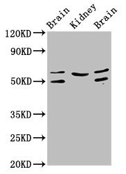
ApplicationsFunctional Assay, Flow Cytometry, ImmunoHistoChemistry, ImmunoHistoChemistry Frozen
Product group Antibodies
ReactivityBovine, Mouse
TargetANGPT2
Overview
- SupplierBiotium
- Product NameVEGF-R2 / CD309 / Flk-1 / KDR3(DC101), Biotin conjugate, 0.1mg/mL [26628-22-8]
- Delivery Days Customer9
- ApplicationsFunctional Assay, Flow Cytometry, ImmunoHistoChemistry, ImmunoHistoChemistry Frozen
- CAS Number26628-22-8
- CertificationResearch Use Only
- ClonalityMonoclonal
- Clone IDDC101
- Concentration0.1 mg/ml
- ConjugateBiotin
- Gene ID285
- Target nameANGPT2
- Target descriptionangiopoietin 2
- Target synonymsAGPT2, ANG2, LMPHM10, angiopoietin-2, Tie2-ligand, angiopoietin-2B, angiopoietin-2a
- HostMouse
- IsotypeIgG1
- Scientific DescriptionThis MAb is specific to Mouse VEGFR2/FLK-1/CD309 and does not cross-react with FLK-2. VEGFR2 is a type I transmembrane glycoprotein. It is a member of the CSF-1/PDGF receptor family of type III tyrosine kinase receptors. Endothelial cells, embryonic tissues, and megakaryocytes mainly express VEGFR2. It plays an important role in the regulation of angiogenesis, vasculogenesis, and vascular permeability. The ligands of VEGFR2 include VEGF-A, VEGF-C, VEGF-D, and VEGF splice isoforms. Ligation of VEGFR2 with its ligands results in the receptor dimerization and auto-phosphorylation, stimulating endothelial cell proliferation and migration. Primary antibodies are available purified, or with a selection of fluorescent CF® Dyes and other labels. CF® Dyes offer exceptional brightness and photostability. Note: Conjugates of blue fluorescent dyes like CF®405S and CF®405M are not recommended for detecting low abundance targets, because blue dyes have lower fluorescence and can give higher non-specific background than other dye colors.
- SourceAnimal
- ReactivityBovine, Mouse
- Storage Instruction2°C to 8°C,RT
- UNSPSC41116161
MSDS
Related products
Product group Antibodies
Anti-ANGPT2 AntibodyA28554
ApplicationsWestern Blot, ImmunoHistoChemistry
ReactivityHuman, Mouse, Rat
- SizePrice
Product group Antibodies
ApplicationsELISA, Neutralisation/Blocking
ReactivityHuman
TargetANGPT2
- SizePrice
Product group Antibodies
Anti-ANGPT2 Antibody144-00698
ApplicationsImmunoFluorescence, Western Blot
ReactivityHuman, Mouse, Rat
TargetANGPT2
- SizePrice
Product group Antibodies

ANGPT2 / Angiopoietin-2 AntibodyLS-C831585
ApplicationsImmunoHistoChemistry
ReactivityMouse, Rat
TargetANGPT2
- SizePrice

Product group Antibodies
Anti-Angiopoietin-2/ANGPT2 Antibody Picoband(r)A00370-2-CARRIER-FREE
ApplicationsFlow Cytometry, ImmunoFluorescence, Western Blot, ELISA, ImmunoCytoChemistry, ImmunoHistoChemistry
ReactivityHuman, Mouse, Rat
TargetANGPT2
- SizePrice
Product group Antibodies
ApplicationsImmunoFluorescence, Western Blot, ELISA, ImmunoCytoChemistry, ImmunoHistoChemistry, ImmunoHistoChemistry Frozen, ImmunoHistoChemistry Paraffin
ReactivityCanine, Chicken, Human, Mouse, Porcine, Rat
TargetANGPT2
- SizePrice
Product group Antibodies
ANGPT2 Polyclonal AntibodyCAC14082
ApplicationsWestern Blot, ELISA, ImmunoHistoChemistry
ReactivityMouse, Rat
TargetANGPT2
- SizePrice
Product group Antibodies
ANGPT2 AntibodyCSB-PA05749A0RB
ApplicationsWestern Blot, ELISA, ImmunoHistoChemistry
ReactivityHuman, Mouse, Rat
TargetANGPT2
- SizePrice
Product group Antibodies
Angiopoietin 2 antibodyGTX100928
ApplicationsWestern Blot
ReactivityHuman, Mouse
TargetANGPT2
- SizePrice