

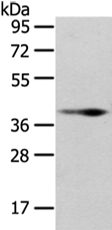
Non-transfected (–) and transfected (+) 293T whole cell extracts (30 μg) were separated by 10% SDS-PAGE, and the membrane was blotted with Wnt10b antibody [HL2370] (GTX638576) diluted at 1:5000. The HRP-conjugated anti-rabbit IgG antibody (GTX213110-01) was used to detect the primary antibody.
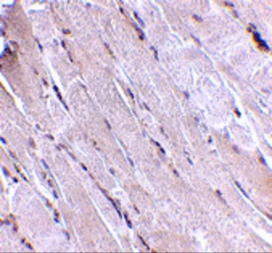
![Wnt10b antibody [HL2370] detects Wnt10b protein by immunohistochemical analysis. Sample: Paraffin-embedded rat tissues. Wnt10b stained by Wnt10b antibody [HL2370] (GTX638576) diluted at 1:100. Antigen Retrieval: Citrate buffer, pH 6.0, 15 min](https://www.genetex.com/upload/website/prouct_img/normal/GTX638576/GTX638576_T-45033_20230512_IHC-P_multiple_R_23060622_854.webp "Wnt10b antibody [HL2370] detects Wnt10b protein by immunohistochemical analysis. Sample: Paraffin-embedded rat tissues. Wnt10b stained by Wnt10b antibody [HL2370] (GTX638576) diluted at 1:100. Antigen Retrieval: Citrate buffer, pH 6.0, 15 min")
![Wnt10b antibody [HL2370] detects Wnt10b protein by immunohistochemical analysis. Sample: Paraffin-embedded mouse tissues. Wnt10b stained by Wnt10b antibody [HL2370] (GTX638576) diluted at 1:100. Antigen Retrieval: Citrate buffer, pH 6.0, 15 min](https://www.genetex.com/upload/website/prouct_img/normal/GTX638576/GTX638576_T-45033_20230512_IHC-P_multiple_M_23060622_236.webp "Wnt10b antibody [HL2370] detects Wnt10b protein by immunohistochemical analysis. Sample: Paraffin-embedded mouse tissues. Wnt10b stained by Wnt10b antibody [HL2370] (GTX638576) diluted at 1:100. Antigen Retrieval: Citrate buffer, pH 6.0, 15 min")
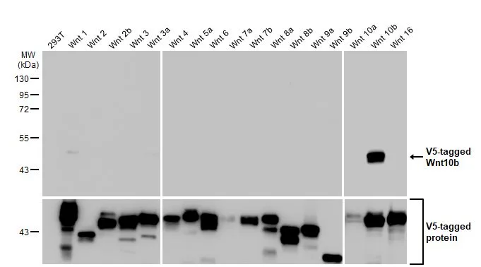
![Various whole cell extracts (30 μg) were separated by 10% SDS-PAGE, and the membrane was blotted with Wnt10b antibody [HL2370] (GTX638576) diluted at 1:500. The HRP-conjugated anti-rabbit IgG antibody (GTX213110-01) was used to detect the primary antibody, and the signal was developed with Trident ECL plus-Enhanced.](https://www.genetex.com/upload/website/prouct_img/normal/GTX638576/GTX638576_45110_20230818_WB_23083020_812.webp "Various whole cell extracts (30 μg) were separated by 10% SDS-PAGE, and the membrane was blotted with Wnt10b antibody [HL2370] (GTX638576) diluted at 1:500. The HRP-conjugated anti-rabbit IgG antibody (GTX213110-01) was used to detect the primary antibody, and the signal was developed with Trident ECL plus-Enhanced.")
![Whole zebrafish extract (30 μg) was separated by 10% SDS-PAGE, and the membrane was blotted with Wnt10b antibody [HL2370] (GTX638576) diluted at 1:1000. The HRP-conjugated anti-rabbit IgG antibody (GTX213110-01) was used to detect the primary antibody, and the signal was developed with Trident ECL plus-Enhanced.](https://www.genetex.com/upload/website/prouct_img/normal/GTX638576/GTX638576_45110_20230908_WB_Z_23091319_577.webp "Whole zebrafish extract (30 μg) was separated by 10% SDS-PAGE, and the membrane was blotted with Wnt10b antibody [HL2370] (GTX638576) diluted at 1:1000. The HRP-conjugated anti-rabbit IgG antibody (GTX213110-01) was used to detect the primary antibody, and the signal was developed with Trident ECL plus-Enhanced.")
![Wnt10b antibody [HL2370] detects Wnt10b protein at cell membrane by immunofluorescent analysis. Sample: HCT116 cells were fixed in 4% paraformaldehyde at RT for 15 min. Green: Wnt10b stained by Wnt10b antibody [HL2370] (GTX638576) diluted at 1:500. Blue: Fluoroshield with DAPI (GTX30920).](https://www.genetex.com/upload/website/prouct_img/normal/GTX638576/GTX638576_45110_20240301_ICC_IF_24030600_741.webp "Wnt10b antibody [HL2370] detects Wnt10b protein at cell membrane by immunofluorescent analysis. Sample: HCT116 cells were fixed in 4% paraformaldehyde at RT for 15 min. Green: Wnt10b stained by Wnt10b antibody [HL2370] (GTX638576) diluted at 1:500. Blue: Fluoroshield with DAPI (GTX30920).")
Non-transfected (–) and transfected (+) 293T whole cell extracts (30 μg) were separated by 10% SDS-PAGE, and the membrane was blotted with Wnt10b antibody [HL2370] (GTX638576) diluted at 1:5000. The HRP-conjugated anti-rabbit IgG antibody (GTX213110-01) was used to detect the primary antibody.
Wnt10b antibody [HL2370]
GTX638576
ApplicationsImmunoFluorescence, Western Blot, ImmunoCytoChemistry, ImmunoHistoChemistry, ImmunoHistoChemistry Paraffin
Product group Antibodies
ReactivityHuman, Mouse, Rat, Zebra Fish
TargetWNT10B
Overview
- SupplierGeneTex
- Product NameWnt10b antibody [HL2370]
- Delivery Days Customer9
- Application Supplier NoteWB: 1:500-1:10000. *Optimal dilutions/concentrations should be determined by the researcher.Not tested in other applications.
- ApplicationsImmunoFluorescence, Western Blot, ImmunoCytoChemistry, ImmunoHistoChemistry, ImmunoHistoChemistry Paraffin
- CertificationResearch Use Only
- ClonalityMonoclonal
- Clone IDHL2370
- Concentration1 mg/ml
- ConjugateUnconjugated
- Gene ID7480
- Target nameWNT10B
- Target descriptionWnt family member 10B
- Target synonymsSHFM6, STHAG8, WNT-12, protein Wnt-10b, WNT-10B protein, wingless-type MMTV integration site family, member 10B
- HostRabbit
- IsotypeIgG
- Protein IDO00744
- Protein NameProtein Wnt-10b
- Scientific DescriptionThe WNT gene family consists of structurally related genes which encode secreted signaling proteins. These proteins have been implicated in oncogenesis and in several developmental processes, including regulation of cell fate and patterning during embryogenesis. This gene is a member of the WNT gene family. It may be involved in breast cancer, and its protein signaling is likely a molecular switch that governs adipogenesis. This protein is 96% identical to the mouse Wnt10b protein at the amino acid level. This gene is clustered with another family member, WNT1, in the chromosome 12q13 region. [provided by RefSeq, Jul 2008]
- ReactivityHuman, Mouse, Rat, Zebra Fish
- Storage Instruction-20°C or -80°C,2°C to 8°C
- UNSPSC41116161
Datasheet
Related products

Product group Antibodies
Anti-WNT10B Antibody Picoband(r)A02574-1-CARRIER-FREE
ApplicationsFlow Cytometry, Western Blot
ReactivityHuman, Mouse, Rat
TargetWNT10B
- SizePrice

Product group Antibodies
References
WNT10B Polyclonal AntibodyBS-3662R
ApplicationsImmunoFluorescence, Western Blot, ELISA, ImmunoCytoChemistry, ImmunoHistoChemistry, ImmunoHistoChemistry Frozen, ImmunoHistoChemistry Paraffin
ReactivityBovine, Canine, Equine, Human, Mouse, Porcine, Rabbit, Rat
TargetWNT10B
- SizePrice
Product group Antibodies
ApplicationsWestern Blot, ImmunoHistoChemistry
ReactivityMouse, Rat
TargetWNT10B
- SizePrice

Product group Antibodies
WNT10B AntibodyCSB-PA164068
ApplicationsWestern Blot, ELISA
ReactivityHuman, Mouse
TargetWNT10B
- SizePrice
Product group Antibodies
References
Wnt10b antibodyGTX17006
ApplicationsWestern Blot, ELISA, ImmunoHistoChemistry, ImmunoHistoChemistry Paraffin
ReactivityHuman, Mouse, Rat
TargetWNT10B
- SizePrice
Product group Antibodies

WNT10B Antibody (aa30-322)LS-C315040
ApplicationsWestern Blot
ReactivityHuman
TargetWNT10B
- SizePrice
![ICC/IF analysis of PANC-1 cells using GTX83307 WNT10B antibody [5A7]. Green : WNT10B Blue: DRAQ5 fluorescent DNA dye Red: Actin filaments](https://www.genetex.com/upload/website/prouct_img/normal/GTX83307/GTX83307_20170912_ICCIF_w_23061322_617.webp)
Product group Antibodies
References
Wnt10b antibody [5A7]GTX83307
ApplicationsImmunoFluorescence, Western Blot, ELISA, ImmunoCytoChemistry, ImmunoHistoChemistry, ImmunoHistoChemistry Paraffin
ReactivityHuman
TargetWNT10B
- SizePrice

Product group Antibodies
Anti-WNT10B AntibodyHPA055048
ApplicationsWestern Blot, ImmunoHistoChemistry
ReactivityHuman
TargetWNT10B
- SizePrice