

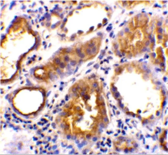
IHC-P analysis of human kidney tissue using GTX31680 ACE2 antibody. Working concentration : 2 μg/ml

 0.5, (B) 1, and (C) 2 μg/ml")
IHC-P analysis of human kidney tissue using GTX31680 ACE2 antibody. Working concentration : 2 μg/ml
ACE2 antibody
GTX31680
ApplicationsWestern Blot, ELISA, ImmunoHistoChemistry, ImmunoHistoChemistry Paraffin
Product group Antibodies
ReactivityHuman, Mouse
TargetACE2
Overview
- SupplierGeneTex
- Product NameACE2 antibody
- Delivery Days Customer9
- Application Supplier NoteWB: 0.5 - 2 microg/mL. IHC-P: 2 microg/mL. *Optimal dilutions/concentrations should be determined by the researcher.Not tested in other applications.
- ApplicationsWestern Blot, ELISA, ImmunoHistoChemistry, ImmunoHistoChemistry Paraffin
- CertificationResearch Use Only
- ClonalityPolyclonal
- Concentration1 mg/ml
- ConjugateUnconjugated
- Gene ID59272
- Target nameACE2
- Target descriptionangiotensin converting enzyme 2
- Target synonymsACEH, angiotensin-converting enzyme 2, ACE-related carboxypeptidase, angiotensin I converting enzyme (peptidyl-dipeptidase A) 2, angiotensin I converting enzyme 2, angiotensin-converting enzyme homolog, angiotensin-converting enzyme-related carboxypeptidase, metalloprotease MPROT15, peptidyl-dipeptidase A
- HostRabbit
- IsotypeIgG
- Protein IDQ9BYF1
- Protein NameAngiotensin-converting enzyme 2
- Scientific DescriptionThe protein encoded by this gene belongs to the angiotensin-converting enzyme family of dipeptidyl carboxydipeptidases and has considerable homology to human angiotensin 1 converting enzyme. This secreted protein catalyzes the cleavage of angiotensin I into angiotensin 1-9, and angiotensin II into the vasodilator angiotensin 1-7. The organ- and cell-specific expression of this gene suggests that it may play a role in the regulation of cardiovascular and renal function, as well as fertility. In addition, the encoded protein is a functional receptor for the spike glycoprotein of the human coronaviruses SARS and HCoV-NL63. [provided by RefSeq, Jul 2008]
- ReactivityHuman, Mouse
- Storage Instruction-20°C or -80°C,2°C to 8°C
- UNSPSC12352203
Datasheet
Related products
Product group Antibodies

Anti-ACE2 [h11B11]AB03703-1.1
ApplicationsFlow Cytometry, ELISA, Neutralisation/Blocking
ReactivityHuman, Monkey
TargetACE2
- SizePrice

Product group Antibodies
Anti-ACE2 AntibodyAMAB91259
ApplicationsWestern Blot, ImmunoHistoChemistry
ReactivityHuman
TargetACE2
- SizePrice
Product group Antibodies
Anti-ACE2 Antibody130-10857
ApplicationsELISA
ReactivityVirus
TargetACE2
- SizePrice

Product group Antibodies
Anti-ACE2 Antibody Picoband(r)A00756-3-CARRIER-FREE
ApplicationsFlow Cytometry, Western Blot, ELISA
ReactivityHuman
TargetACE2
- SizePrice
![ACE2 antibody [SN0754] detects ACE2 protein at cell membrane by immunohistochemical analysis. Sample: Paraffin-embedded mouse kidney. ACE2 stained by ACE2 antibody [SN0754] (GTX01160) diluted at 1:2000. Antigen Retrieval: Citrate buffer, pH 6.0, 15 min](https://www.genetex.com/upload/website/prouct_img/normal/GTX01160/GTX01160_HK0921_20200313_IHC-P_M_w_23053121_166.webp)
Product group Antibodies
References
ACE2 antibody [SN0754]GTX01160
ApplicationsImmunoFluorescence, Western Blot, ImmunoCytoChemistry, ImmunoHistoChemistry, ImmunoHistoChemistry Paraffin
ReactivityHuman, Monkey, Mouse, Rat
TargetACE2
- SizePrice
![IHC-P analysis of human testis tissue using GTX04425 ACE2 antibody [MSVA-919R] HistoMAX?. Leydig cells and spermatocytes of the testis show a particularly strong ACE2 immunostaining in the testis.](https://www.genetex.com/upload/website/prouct_img/normal/GTX04425/GTX04425_20230728_IHC-P_2_23072722_840.webp)
Product group Antibodies
ApplicationsImmunoHistoChemistry, ImmunoHistoChemistry Paraffin
ReactivityHuman
TargetACE2
- SizePrice
![Mouse tissue extract (50 μg) was separated by 7.5% SDS-PAGE, and the membrane was blotted with ACE2 antibody [N1N2], N-term (GTX101395) diluted at 1:500. The HRP-conjugated anti-rabbit IgG antibody (GTX213110-01) was used to detect the primary antibody, and the signal was developed with Trident ECL plus-Enhanced.](https://www.genetex.com/upload/website/prouct_img/normal/GTX101395/GTX101395_43824_20200327_WB_M_kidney_w_23060100_326.webp)
Product group Antibodies
References
ACE2 antibody [N1N2], N-termGTX101395
ApplicationsFlow Cytometry, ImmunoFluorescence, Western Blot, ELISA, ImmunoCytoChemistry, ImmunoHistoChemistry, ImmunoHistoChemistry Paraffin
ReactivityHuman, Monkey, Mouse, Rat
TargetACE2
- SizePrice
![Whole cell extract (30 μg) was separated by 7.5% SDS-PAGE, and the membrane was blotted with ACE2 antibody [GT19410] (GTX635897) diluted at 1:1000. The HRP-conjugated anti-mouse IgG antibody (GTX213111-01) was used to detect the primary antibody.](https://www.genetex.com/upload/website/prouct_img/normal/GTX635897/GTX635897_44979_20230317_WB_Monkey_23032819_299.webp)
Product group Antibodies
ACE2 antibody [GT19410]GTX635897
ApplicationsImmunoFluorescence, Western Blot, ELISA, ImmunoCytoChemistry, ImmunoHistoChemistry, ImmunoHistoChemistry Paraffin, Other Application
ReactivityHuman, Monkey, Mouse
TargetACE2
- SizePrice
![Non-transfected (–) and transfected (+) 293T whole cell extracts (30 μg) were separated by 7.5% SDS-PAGE, and the membrane was blotted with ACE2 antibody [HL1092] (GTX636265) diluted at 1:5000. The HRP-conjugated anti-rabbit IgG antibody (GTX213110-01) was used to detect the primary antibody.](https://www.genetex.com/upload/website/prouct_img/normal/GTX636265/GTX636265_44389_20220812_WB_B_22081423_460.webp)
Product group Antibodies
ACE2 antibody [HL1092]GTX636265
ApplicationsImmunoFluorescence, Western Blot, ImmunoCytoChemistry, ImmunoHistoChemistry, ImmunoHistoChemistry Paraffin
ReactivityCanine, Feline, Human, Monkey
TargetACE2
- SizePrice