

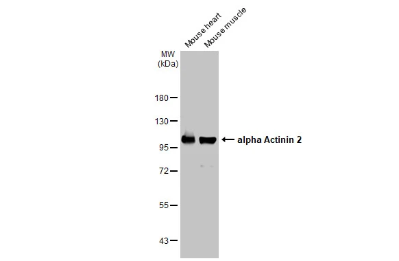
Mouse tissue extract (50 μg) were separated by 7.5% SDS-PAGE, and the membrane was blotted with alpha Actinin 2 antibody [N2C1], Internal (GTX111167) diluted at 1:1000. The HRP-conjugated anti-rabbit IgG antibody (GTX213110-01) was used to detect the primary antibody.
![Immunohistochemical analysis of paraffin-embedded zebrafish tissue, using alpha Actinin 2 antibody [N2C1], Internal (GTX111167) at 1:300 dilution.](https://www.genetex.com/upload/website/prouct_img/normal/GTX111167/GTX111167_40086_IHC_Z_22111423_487.webp "Immunohistochemical analysis of paraffin-embedded zebrafish tissue, using alpha Actinin 2 antibody [N2C1], Internal (GTX111167) at 1:300 dilution.")
 A: adult zebrafish 7.5% SDS PAGE GTX111167 diluted at 1:3000")
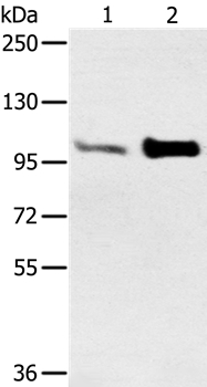
 A: A431 (GTX27909) B: H1299 7.5% SDS PAGE GTX111167 diluted at 1:1000")
 was separated by 7.5% SDS-PAGE, and the membrane was blotted with alpha Actinin 2 antibody (GTX111167) diluted at 1:5000.")
 antibody at 1:200 dilution.")
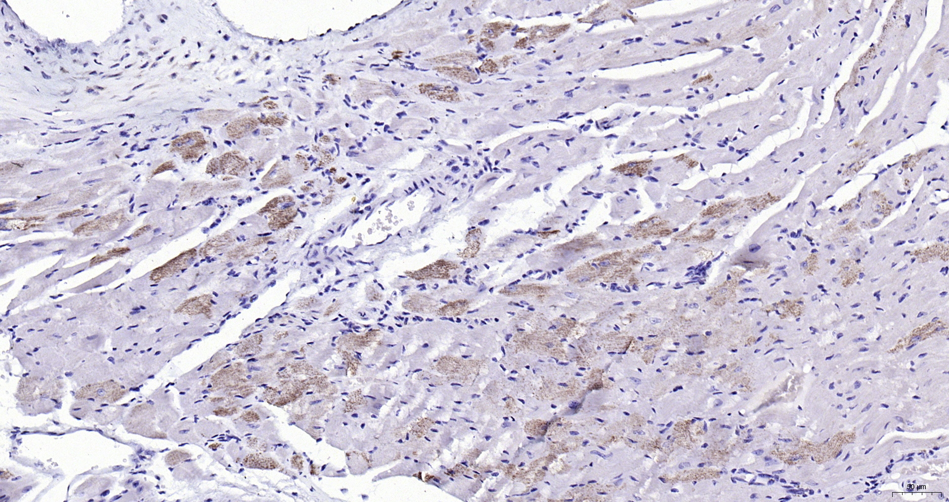
![alpha Actinin 2 antibody [N2C1], Internal detects alpha Actinin 2 protein at cytoplasm and membrane on human breast carcinoma by immunohistochemical analysis. Sample: Paraffin-embedded human breast carcinoma. alpha Actinin 2 antibody [N2C1], Internal (GTX111167) diluted at 1:250.
Antigen Retrieval: Trilogy? (EDTA based, pH 8.0) buffer, 15min](https://www.genetex.com/upload/website/prouct_img/normal/GTX111167/GTX111167_40086_20141128_IHC_w_23060500_965.webp "alpha Actinin 2 antibody [N2C1], Internal detects alpha Actinin 2 protein at cytoplasm and membrane on human breast carcinoma by immunohistochemical analysis. Sample: Paraffin-embedded human breast carcinoma. alpha Actinin 2 antibody [N2C1], Internal (GTX111167) diluted at 1:250.
Antigen Retrieval: Trilogy? (EDTA based, pH 8.0) buffer, 15min")
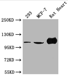
 A: NIH-3T3 7.5% SDS PAGE GTX111167 diluted at 1:3000")
Mouse tissue extract (50 μg) were separated by 7.5% SDS-PAGE, and the membrane was blotted with alpha Actinin 2 antibody [N2C1], Internal (GTX111167) diluted at 1:1000. The HRP-conjugated anti-rabbit IgG antibody (GTX213110-01) was used to detect the primary antibody.
alpha Actinin 2 antibody [N2C1], Internal
GTX111167
ApplicationsImmunoFluorescence, Western Blot, ImmunoCytoChemistry, ImmunoHistoChemistry, ImmunoHistoChemistry Paraffin
Product group Antibodies
ReactivityHuman, Mouse, Rat, Zebra Fish
TargetACTN2
Overview
- SupplierGeneTex
- Product Namealpha Actinin 2 antibody [N2C1], Internal
- Delivery Days Customer9
- Application Supplier NoteWB: 1:500-1:10000. ICC/IF: 1:100-1:1000. IHC-P: 1:100-1:1000. *Optimal dilutions/concentrations should be determined by the researcher.Not tested in other applications.
- ApplicationsImmunoFluorescence, Western Blot, ImmunoCytoChemistry, ImmunoHistoChemistry, ImmunoHistoChemistry Paraffin
- CertificationResearch Use Only
- ClonalityPolyclonal
- Concentration1 mg/ml
- ConjugateUnconjugated
- Gene ID88
- Target nameACTN2
- Target descriptionactinin alpha 2
- Target synonymsCMD1AA, CMH23, CMYO8, CMYP8, MPD6, MYOCOZ, alpha-actinin-2, F-actin cross-linking protein, alpha-actinin skeletal muscle
- HostRabbit
- IsotypeIgG
- Protein IDP35609
- Protein NameAlpha-actinin-2
- Scientific DescriptionAlpha actinins belong to the spectrin gene superfamily which represents a diverse group of cytoskeletal proteins, including the alpha and beta spectrins and dystrophins. Alpha actinin is an actin-binding protein with multiple roles in different cell types. In nonmuscle cells, the cytoskeletal isoform is found along microfilament bundles and adherens-type junctions, where it is involved in binding actin to the membrane. In contrast, skeletal, cardiac, and smooth muscle isoforms are localized to the Z-disc and analogous dense bodies, where they help anchor the myofibrillar actin filaments. This gene encodes a muscle-specific, alpha actinin isoform that is expressed in both skeletal and cardiac muscles. [provided by RefSeq]
- ReactivityHuman, Mouse, Rat, Zebra Fish
- Storage Instruction-20°C or -80°C,2°C to 8°C
- UNSPSC41116161
Datasheet
Related products
Product group Antibodies
Anti-ACTN2 AntibodyA38104
ApplicationsWestern Blot, ImmunoHistoChemistry
ReactivityHuman, Mouse
- SizePrice

Product group Antibodies
Anti-ACTN2 Antibody Picoband(r)A03673-1-CARRIER-FREE
ApplicationsFlow Cytometry, Western Blot, ELISA, ImmunoHistoChemistry
ReactivityHuman, Mouse, Rat
TargetACTN2
- SizePrice
Product group Antibodies
Anti-ACTN2 Antibody144-03718
ApplicationsImmunoFluorescence, Western Blot, ImmunoHistoChemistry
ReactivityHuman, Mouse, Rat
TargetACTN2
- SizePrice
Product group Antibodies
References
ApplicationsImmunoFluorescence, Western Blot, ELISA, ImmunoCytoChemistry, ImmunoHistoChemistry, ImmunoHistoChemistry Frozen, ImmunoHistoChemistry Paraffin
ReactivityBovine, Canine, Chicken, Equine, Human, Mouse, Porcine, Rabbit, Rat, Sheep
TargetACTN2
- SizePrice
Product group Antibodies
ACTN2 AntibodyCSB-PA001242LA01HU
ApplicationsWestern Blot, ELISA, ImmunoHistoChemistry
ReactivityHuman, Rat
TargetACTN2
- SizePrice
Product group Antibodies
ACTN2 Polyclonal AntibodyCAC15868
ApplicationsWestern Blot, ELISA, ImmunoHistoChemistry
ReactivityRat
TargetACTN2
- SizePrice
Product group Antibodies

ACTN2 AntibodyLS-C403508
ApplicationsWestern Blot, ELISA, ImmunoHistoChemistry
ReactivityHuman, Mouse
TargetACTN2
- SizePrice
![Various whole cell extracts (30 μg) were separated by 5% SDS-PAGE, and the membrane was blotted with alpha Actinin 2 antibody [N1N3] (GTX103219) diluted at 1:1000. The HRP-conjugated anti-rabbit IgG antibody (GTX213110-01) was used to detect the primary antibody.](https://www.genetex.com/upload/website/prouct_img/normal/GTX103219/GTX103219_44335_20210702_WB_M_22072519_433.webp)
Product group Antibodies
alpha Actinin 2 antibody [N1N3]GTX103219
ApplicationsImmunoFluorescence, Western Blot, ImmunoCytoChemistry, ImmunoHistoChemistry, ImmunoHistoChemistry Paraffin
ReactivityHuman, Mouse, Porcine, Rat, Zebra Fish
TargetACTN2
- SizePrice

Product group Antibodies
Anti-ACTN2 AntibodyHPA008315
ApplicationsImmunoHistoChemistry
ReactivityHuman
TargetACTN2
- SizePrice