


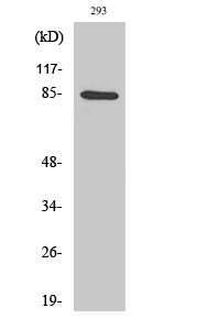
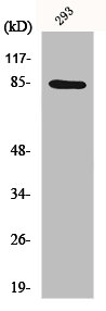
Figure 1. Western blot analysis of IKK alpha using anti-IKK alpha antibody (PB9110). Electrophoresis was performed on a 5-20% SDS-PAGE gel at 70V (Stacking gel) / 90V (Resolving gel) for 2-3 hours. The sample well of each lane was loaded with 30 ug of sample under reducing conditions. Lane 1: human placenta tissue lysates, Lane 2: human K562 whole cell lysates, Lane 3: human HepG2 whole cell lysates, Lane 4: human Caco-2 whole cell lysates. After electrophoresis, proteins were transferred to a nitrocellulose membrane at 150 mA for 50-90 minutes. Blocked the membrane with 5% non-fat milk/TBS for 1.5 hour at RT. The membrane was incubated with rabbit anti-IKK alpha antigen affinity purified polyclonal antibody (Catalog # PB9110) at 0.5 microg/mL overnight at 4°C, then washed with TBS-0.1%Tween 3 times with 5 minutes each and probed with a goat anti-rabbit IgG-HRP secondary antibody at a dilution of 1:5000 for 1.5 hour at RT. The signal is developed using an Enhanced Chemiluminescent detection (ECL) kit (Catalog # EK1002) with Tanon 5200 system. A specific band was detected for IKK alpha at approximately 85 kDa. The expected band size for IKK alpha is at 85 kDa.
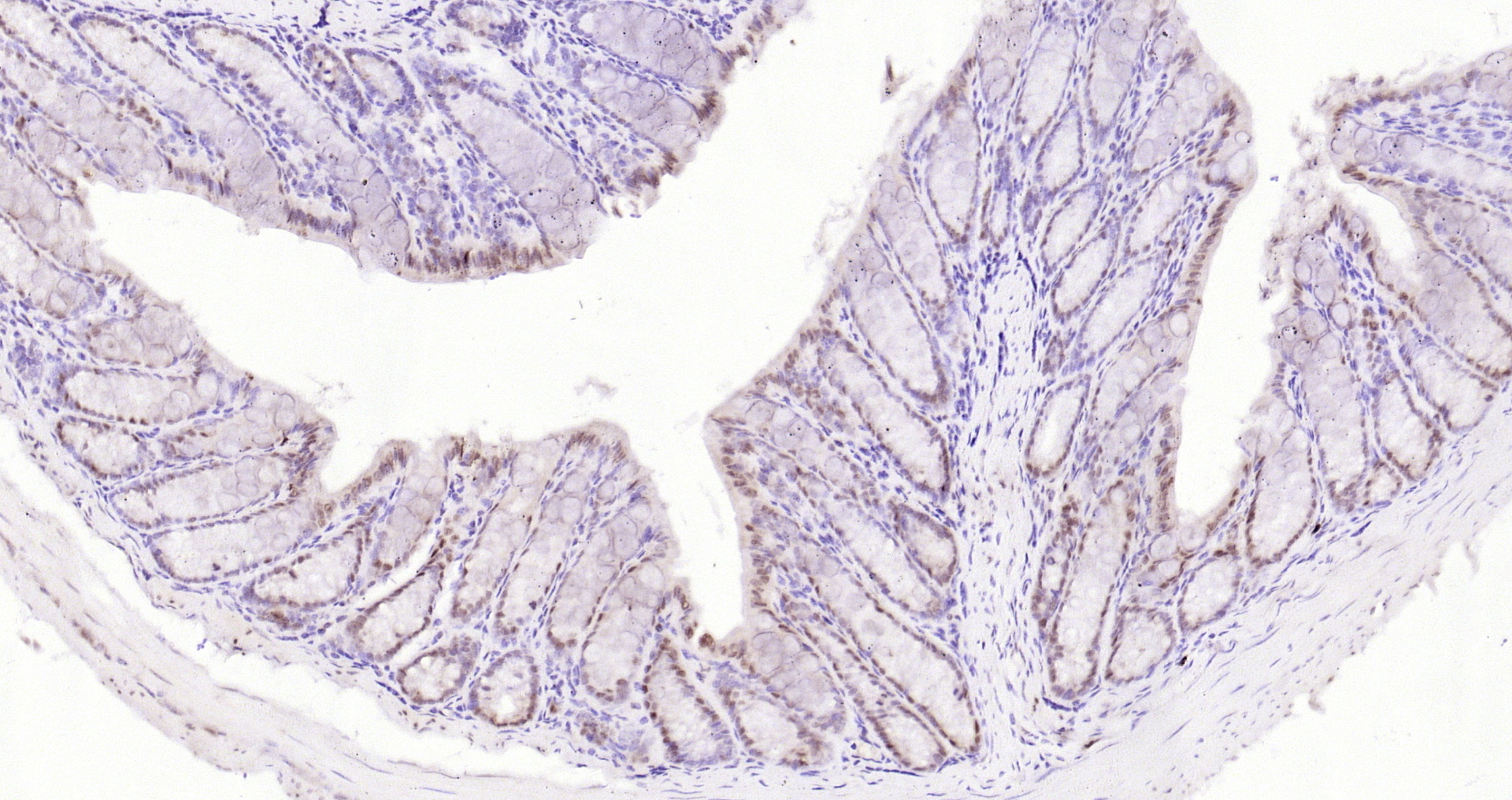
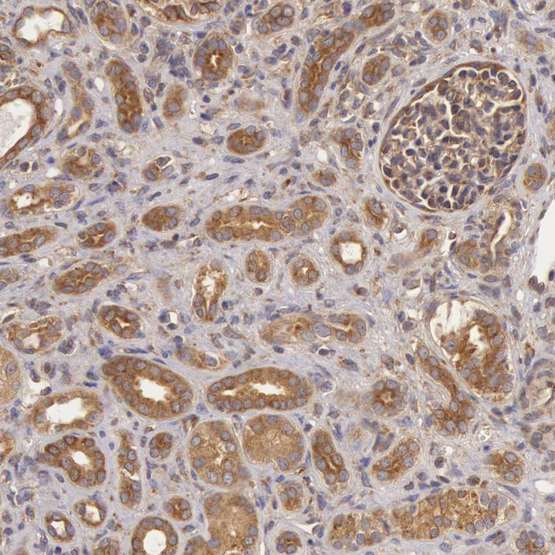
. IKKA was detected in paraffin-embedded section of human lung cancer tissues. Heat mediated antigen retrieval was performed in citrate buffer (pH6, epitope retrieval solution) for 20 mins. The tissue section was blocked with 10% goat serum. The tissue section was then incubated with 1microg/ml rabbit anti-IKKA Antibody (PB9110) overnight at 4°C. Biotinylated goat anti-rabbit IgG was used as secondary antibody and incubated for 30 minutes at 37°C. The tissue section was developed using Strepavidin-Biotin-Complex (SABC)(Catalog # SA1022) with DAB as the chromogen.")
Figure 1. Western blot analysis of IKK alpha using anti-IKK alpha antibody (PB9110). Electrophoresis was performed on a 5-20% SDS-PAGE gel at 70V (Stacking gel) / 90V (Resolving gel) for 2-3 hours. The sample well of each lane was loaded with 30 ug of sample under reducing conditions. Lane 1: human placenta tissue lysates, Lane 2: human K562 whole cell lysates, Lane 3: human HepG2 whole cell lysates, Lane 4: human Caco-2 whole cell lysates. After electrophoresis, proteins were transferred to a nitrocellulose membrane at 150 mA for 50-90 minutes. Blocked the membrane with 5% non-fat milk/TBS for 1.5 hour at RT. The membrane was incubated with rabbit anti-IKK alpha antigen affinity purified polyclonal antibody (Catalog # PB9110) at 0.5 microg/mL overnight at 4°C, then washed with TBS-0.1%Tween 3 times with 5 minutes each and probed with a goat anti-rabbit IgG-HRP secondary antibody at a dilution of 1:5000 for 1.5 hour at RT. The signal is developed using an Enhanced Chemiluminescent detection (ECL) kit (Catalog # EK1002) with Tanon 5200 system. A specific band was detected for IKK alpha at approximately 85 kDa. The expected band size for IKK alpha is at 85 kDa.
Anti-IKK Alpha Picoband Antibody
PB9110
ApplicationsWestern Blot, ImmunoHistoChemistry
Product group Antibodies
ReactivityHuman
TargetCHUK
Overview
- SupplierBoster Bio
- Product NameAnti-IKK Alpha Picoband Antibody
- Delivery Days Customer9
- Application Supplier NoteWB: The detection limit for IKK alpha is approximately 0.2ng/lane under reducing conditions. Tested Species: In-house tested species with positive results. By Heat: Boiling the paraffin sections in 10mM citrate buffer, pH6.0, for 20mins is required for the staining of formalin/paraffin sections. Other applications have not been tested. Optimal dilutions should be determined by end users.
- ApplicationsWestern Blot, ImmunoHistoChemistry
- Applications SupplierIHP, WB, IHC
- CertificationResearch Use Only
- ClonalityPolyclonal
- Concentration500 ug/ml
- Gene ID1147
- Target nameCHUK
- Target descriptioncomponent of inhibitor of nuclear factor kappa B kinase complex
- Target synonymsBPS2, IKBKA, IKK-1, IKK-alpha, IKK1, IKKA, NFKBIKA, TCF16, inhibitor of nuclear factor kappa-B kinase subunit alpha, I-kappa-B kinase 1, I-kappa-B kinase-alpha, IKK-a kinase, IkB kinase alpha subunit, Nuclear factor NFkappaB inhibitor kinase alpha, conserved helix-loop-helix ubiquitous kinase, transcription factor 16
- HostRabbit
- IsotypeIgG
- Protein IDO15111
- Protein NameInhibitor of nuclear factor kappa-B kinase subunit alpha
- Scientific DescriptionBoster Bio Anti-IKK alpha/CHUK Antibody Picoband® catalog # PB9110. Tested in IHC, WB applications. This antibody reacts with Human. The brand Picoband indicates this is a premium antibody that guarantees superior quality, high affinity, and strong signals with minimal background in Western blot applications. Only our best-performing antibodies are designated as Picoband, ensuring unmatched performance.
- ReactivityHuman
- Reactivity SupplierHuman, Mouse, Rat
- Storage Instruction-20°C,2°C to 8°C
- UNSPSC12352203
Datasheet
MSDS
Related products
Product group Antibodies
IKK alpha Polyclonal AntibodyBS-2907R
ApplicationsImmunoFluorescence, ELISA, ImmunoCytoChemistry, ImmunoHistoChemistry, ImmunoHistoChemistry Frozen, ImmunoHistoChemistry Paraffin
ReactivityCanine, Equine, Human, Mouse, Rat
TargetCHUK
- SizePrice
Product group Antibodies
Anti-CHUK Antibody144-02062
ApplicationsImmunoFluorescence, ImmunoPrecipitation, Western Blot, ImmunoHistoChemistry
ReactivityHuman, Mouse, Rat
TargetCHUK
- SizePrice
Product group Antibodies
Anti-IKKalpha AntibodyA36019
ApplicationsWestern Blot, ELISA, ImmunoHistoChemistry
ReactivityHuman, Mouse, Rat
- SizePrice
Product group Antibodies

Goat anti-CHUK AntibodyEB11382
ApplicationsWestern Blot, ELISA
ReactivityHuman
TargetCHUK
- SizePrice
Product group Antibodies
References
IKK alpha antibodyGTX132964
ApplicationsWestern Blot
ReactivityHuman
TargetCHUK
- SizePrice
Product group Antibodies

CHUK / IKKA / IKK Alpha AntibodyLS-C746736
ApplicationsImmunoFluorescence, Western Blot
ReactivityHuman, Mouse, Rat
TargetCHUK
- SizePrice
Product group Antibodies
Anti-CHUK AntibodyHPA001402
ApplicationsImmunoCytoChemistry, ImmunoHistoChemistry
ReactivityHuman
TargetCHUK
- SizePrice
Product group Antibodies
CHUK AntibodyCSB-PA003008
ApplicationsWestern Blot, ELISA, ImmunoHistoChemistry
ReactivityHuman, Mouse, Rat
TargetCHUK
- SizePrice