


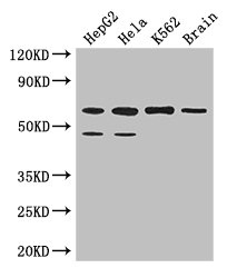
Figure 1. Western blot analysis of PAF1 using anti-PAF1 antibody (A01640-1). Electrophoresis was performed on a 5-20% SDS-PAGE gel at 70V (Stacking gel) / 90V (Resolving gel) for 2-3 hours. The sample well of each lane was loaded with 50ug of sample under reducing conditions. Lane 1: human A549 whole cell lysates, Lane 2: human A431 whole cell lysates, Lane 3: human Caco-2 whole cell lysates, Lane 4: human SW620 whole cell lysates, Lane 5: rat heart tissue lysates, Lane 6: mouse heart tissue lysates. After Electrophoresis, proteins were transferred to a Nitrocellulose membrane at 150mA for 50-90 minutes. Blocked the membrane with 5% Non-fat Milk/ TBS for 1.5 hour at RT. The membrane was incubated with rabbit anti-PAF1 antigen affinity purified polyclonal antibody (Catalog # A01640-1) at 0.25 microg/mL overnight at 4°C, then washed with TBS-0.1%Tween 3 times with 5 minutes each and probed with a goat anti-rabbit IgG-HRP secondary antibody at a dilution of 1:5000 for 1.5 hour at RT. The signal is developed using an Enhanced Chemiluminescent detection (ECL) kit (Catalog # EK1002) with Tanon 5200 system. A specific band was detected for PAF1 at approximately 80KD. The expected band size for PAF1 is at 60KD.
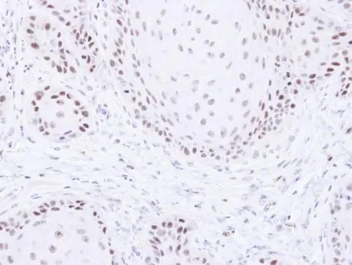
. PAF1 was detected in immunocytochemical section of A549 cells. Enzyme antigen retrieval was performed using IHC enzyme antigen retrieval reagent (AR0022) for 15 mins. The cells were blocked with 10% goat serum. And then incubated with 2microg/mL rabbit anti-PAF1 Antibody (A01640-1) overnight at 4°C. DyLight®488 conjugated Goat Anti-Rabbit IgG (BA1127) was used as secondary antibody at 1:100 dilution and incubated for 30 minutes at 37°C. The section was counterstained with DAPI. Visualize using a fluorescence microscope and filter sets appropriate for the label used.")
Figure 1. Western blot analysis of PAF1 using anti-PAF1 antibody (A01640-1). Electrophoresis was performed on a 5-20% SDS-PAGE gel at 70V (Stacking gel) / 90V (Resolving gel) for 2-3 hours. The sample well of each lane was loaded with 50ug of sample under reducing conditions. Lane 1: human A549 whole cell lysates, Lane 2: human A431 whole cell lysates, Lane 3: human Caco-2 whole cell lysates, Lane 4: human SW620 whole cell lysates, Lane 5: rat heart tissue lysates, Lane 6: mouse heart tissue lysates. After Electrophoresis, proteins were transferred to a Nitrocellulose membrane at 150mA for 50-90 minutes. Blocked the membrane with 5% Non-fat Milk/ TBS for 1.5 hour at RT. The membrane was incubated with rabbit anti-PAF1 antigen affinity purified polyclonal antibody (Catalog # A01640-1) at 0.25 microg/mL overnight at 4°C, then washed with TBS-0.1%Tween 3 times with 5 minutes each and probed with a goat anti-rabbit IgG-HRP secondary antibody at a dilution of 1:5000 for 1.5 hour at RT. The signal is developed using an Enhanced Chemiluminescent detection (ECL) kit (Catalog # EK1002) with Tanon 5200 system. A specific band was detected for PAF1 at approximately 80KD. The expected band size for PAF1 is at 60KD.
Anti-PAF1/PD2 Picoband(r) Antibody
A01640-1-CARRIER-FREE
ApplicationsImmunoFluorescence, Western Blot, ELISA, ImmunoCytoChemistry
Product group Antibodies
ReactivityHuman, Mouse, Rat
TargetPAF1
Overview
- SupplierBoster Bio
- Product NameAnti-PAF1/PD2 Picoband(r) Antibody
- Delivery Days Customer9
- ApplicationsImmunoFluorescence, Western Blot, ELISA, ImmunoCytoChemistry
- CertificationResearch Use Only
- ClonalityPolyclonal
- Concentration500 ug/ml
- Gene ID54623
- Target namePAF1
- Target descriptionPAF1 component of Paf1/RNA polymerase II complex
- Target synonymsF23149_1, PD2, RNA polymerase II-associated factor 1 homolog, PAF1 homolog, Paf1/RNA polymerase II complex component, Paf1, RNA polymerase II associated factor, homolog, pancreatic differentiation protein 2
- HostRabbit
- IsotypeIgG
- Protein IDQ8N7H5
- Protein NameRNA polymerase II-associated factor 1 homolog
- Scientific DescriptionBoster Bio Anti-PAF1/PD2 Picoband® Antibody catalog # A01640-1. Tested in ELISA, IF, ICC, WB applications. This antibody reacts with Human, Mouse, Rat. The brand Picoband indicates this is a premium antibody that guarantees superior quality, high affinity, and strong signals with minimal background in Western blot applications. Only our best-performing antibodies are designated as Picoband, ensuring unmatched performance.
- ReactivityHuman, Mouse, Rat
- Storage Instruction-20°C,2°C to 8°C
- UNSPSC12352203
Related products
Product group Antibodies

PD2 / PAF1 AntibodyLS-C830621
ApplicationsELISA, ImmunoHistoChemistry
ReactivityHuman, Mouse, Rat
TargetPAF1
- SizePrice

Product group Antibodies
Anti-PAF1 AntibodyHPA041875
ApplicationsImmunoHistoChemistry
ReactivityHuman
TargetPAF1
- SizePrice
Product group Antibodies
PAF1 AntibodyCSB-PA843285LA01HU
ApplicationsWestern Blot, ELISA, ImmunoHistoChemistry
ReactivityHuman, Mouse
TargetPAF1
- SizePrice
Product group Antibodies
PAF1 Polyclonal AntibodyCAC14324
ApplicationsWestern Blot, ELISA, ImmunoHistoChemistry
ReactivityMouse
TargetPAF1
- SizePrice
Product group Antibodies
PAF1 antibodyGTX107346
ApplicationsWestern Blot, ImmunoHistoChemistry, ImmunoHistoChemistry Paraffin
ReactivityHuman
TargetPAF1
- SizePrice
Product group Antibodies
PAF1 Recombinant AntibodyBSM-62168R
ApplicationsWestern Blot
ReactivityHuman, Mouse, Rat
TargetPAF1
- SizePrice