

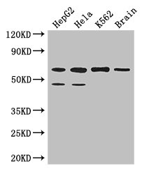
Western Blot Positive WB detected in: HepG2 whole cell lysate, Hela whole cell lysate, K562 whole cell lysate, Mouse brain tissue All lanes: PAF1 antibody at 3.2microg/ml Secondary Goat polyclonal to rabbit IgG at 1/50000 dilution Predicted band size: 60, 56 kDa Observed band size: 60, 48 kDa

Western Blot Positive WB detected in: HepG2 whole cell lysate, Hela whole cell lysate, K562 whole cell lysate, Mouse brain tissue All lanes: PAF1 antibody at 3.2microg/ml Secondary Goat polyclonal to rabbit IgG at 1/50000 dilution Predicted band size: 60, 56 kDa Observed band size: 60, 48 kDa
PAF1 Antibody
CSB-PA843285LA01HU
ApplicationsWestern Blot, ELISA, ImmunoHistoChemistry
Product group Antibodies
ReactivityHuman, Mouse
TargetPAF1
Overview
- SupplierCusabio
- Product NamePAF1 Antibody
- Delivery Days Customer20
- ApplicationsWestern Blot, ELISA, ImmunoHistoChemistry
- CertificationResearch Use Only
- ClonalityPolyclonal
- ConjugateUnconjugated
- Gene ID54623
- Target namePAF1
- Target descriptionPAF1 component of Paf1/RNA polymerase II complex
- Target synonymsF23149_1, PD2, RNA polymerase II-associated factor 1 homolog, PAF1 homolog, Paf1/RNA polymerase II complex component, Paf1, RNA polymerase II associated factor, homolog, pancreatic differentiation protein 2
- HostRabbit
- IsotypeIgG
- Protein IDQ8N7H5
- Protein NameRNA polymerase II-associated factor 1 homolog
- Scientific DescriptionComponent of the PAF1 complex (PAF1C) which has multiple functions during transcription by RNA polymerase II and is implicated in regulation of development and maintenance of embryonic stem cell pluripotency. PAF1C associates with RNA polymerase II through interaction with POLR2A CTD non-phosphorylated and Ser-2- and Ser-5-phosphorylated forms and is involved in transcriptional elongation, acting both indepentently and synergistically with TCEA1 and in cooperation with the DSIF complex and HTATSF1. PAF1C is required for transcription of Hox and Wnt target genes. PAF1C is involved in hematopoiesis and stimulates transcriptional activity of KMT2A/MLL1; it promotes leukemogenesis through association with KMT2A/MLL1-rearranged oncoproteins, such as KMT2A/MLL1-MLLT3/AF9 and KMT2A/MLL1-MLLT1/ENL. PAF1C is involved in histone modifications such as ubiquitination of histone H2B and methylation on histone H3 Lys-4 (H3K4me3). PAF1C recruits the RNF20/40 E3 ubiquitin-protein ligase complex and the E2 enzyme UBE2A or UBE2B to chromatin which mediate monoubiquitination of Lys-120 of histone H2B (H2BK120ub1); UB2A/B-mediated H2B ubiquitination is proposed to be coupled to transcription. PAF1C is involved in mRNA 3 end formation probably through association with cleavage and poly(A) factors. In case of infection by influenza A strain H3N2, PAF1C associates with viral NS1 protein, thereby regulating gene transcription. Connects PAF1C with the RNF20/40 E3 ubiquitin-protein ligase complex. Involved in polyadenylation of mRNA precursors. Has oncogenic activity in vivo and in vitro.
- ReactivityHuman, Mouse
- Storage Instruction-20°C or -80°C
- UNSPSC41116161
Related products

Product group Antibodies
Anti-PAF1/PD2 Picoband(r) AntibodyA01640-1-CARRIER-FREE
ApplicationsImmunoFluorescence, Western Blot, ELISA, ImmunoCytoChemistry
ReactivityHuman, Mouse, Rat
TargetPAF1
- SizePrice
Product group Antibodies

PD2 / PAF1 AntibodyLS-C830621
ApplicationsELISA, ImmunoHistoChemistry
ReactivityHuman, Mouse, Rat
TargetPAF1
- SizePrice

Product group Antibodies
Anti-PAF1 AntibodyHPA041875
ApplicationsImmunoHistoChemistry
ReactivityHuman
TargetPAF1
- SizePrice
Product group Antibodies
PAF1 Polyclonal AntibodyCAC14324
ApplicationsWestern Blot, ELISA, ImmunoHistoChemistry
ReactivityMouse
TargetPAF1
- SizePrice
Product group Antibodies
PAF1 antibodyGTX107346
ApplicationsWestern Blot, ImmunoHistoChemistry, ImmunoHistoChemistry Paraffin
ReactivityHuman
TargetPAF1
- SizePrice
Product group Antibodies
PAF1 Recombinant AntibodyBSM-62168R
ApplicationsWestern Blot
ReactivityHuman, Mouse, Rat
TargetPAF1
- SizePrice