

PAF1 Monoclonal Antibody
CAB3437
ReactivityHuman
Product group Antibodies
TargetPAF1
Overview
- SupplierAssay Genie
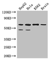
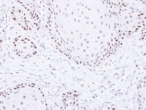
- Product NamePAF1 Monoclonal Antibody
- Delivery Days Customer9
- Applications SupplierWB,IHC
- CertificationResearch Use Only
- ClonalityMonoclonal
- Gene ID54623
- Target namePAF1
- Target descriptionPAF1 component of Paf1/RNA polymerase II complex
- Target synonymsF23149_1, PD2, RNA polymerase II-associated factor 1 homolog, PAF1 homolog, Paf1/RNA polymerase II complex component, Paf1, RNA polymerase II associated factor, homolog, pancreatic differentiation protein 2
- HostRabbit
- Protein IDQ8N7H5
- Protein NameRNA polymerase II-associated factor 1 homolog
- Shelf life instruction12 months
- SourceRabbit
- ReactivityHuman
- Reactivity SupplierHuman,Mouse,Rat
- Storage Instruction-20°C
- UNSPSC12352203
Related products

Product group Antibodies
Anti-PAF1/PD2 Picoband(r) AntibodyA01640-1-CARRIER-FREE
ApplicationsImmunoFluorescence, Western Blot, ELISA, ImmunoCytoChemistry
ReactivityHuman, Mouse, Rat
TargetPAF1
- SizePrice
Product group Antibodies

PD2 / PAF1 AntibodyLS-C830621
ApplicationsELISA, ImmunoHistoChemistry
ReactivityHuman, Mouse, Rat
TargetPAF1
- SizePrice

Product group Antibodies
Anti-PAF1 AntibodyHPA041875
ApplicationsImmunoHistoChemistry
ReactivityHuman
TargetPAF1
- SizePrice
Product group Antibodies
PAF1 AntibodyCSB-PA843285LA01HU
ApplicationsWestern Blot, ELISA, ImmunoHistoChemistry
ReactivityHuman, Mouse
TargetPAF1
- SizePrice
Product group Antibodies
PAF1 Polyclonal AntibodyCAC14324
ApplicationsWestern Blot, ELISA, ImmunoHistoChemistry
ReactivityMouse
TargetPAF1
- SizePrice
Product group Antibodies
PAF1 antibodyGTX107346
ApplicationsWestern Blot, ImmunoHistoChemistry, ImmunoHistoChemistry Paraffin
ReactivityHuman
TargetPAF1
- SizePrice
Product group Antibodies
PAF1 Recombinant AntibodyBSM-62168R
ApplicationsWestern Blot
ReactivityHuman, Mouse, Rat
TargetPAF1
- SizePrice