


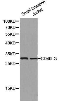
Figure 1. Western blot analysis of TRAP/CD40L/CD40LG using anti-TRAP/CD40L/CD40LG antibody (A01114-4). Electrophoresis was performed on a 5-20% SDS-PAGE gel at 70V (Stacking gel) / 90V (Resolving gel) for 2-3 hours. The sample well of each lane was loaded with 30ug of sample under reducing conditions. Lane 1: human HL-60 whole cell lysates, Lane 2: human K562 whole cell lysates, Lane 3: human Raji whole cell lysates, Lane 4: human Jurkat whole cell lysates, Lane 5: rat RH35 whole cell lysates, Lane 6: mouse spleen tissue lysates, Lane 7: mouse ANA-1 whole cell lysates. After Electrophoresis, proteins were transferred to a Nitrocellulose membrane at 150mA for 50-90 minutes. Blocked the membrane with 5% Non-fat Milk/ TBS for 1.5 hour at RT. The membrane was incubated with rabbit anti-TRAP/CD40L/CD40LG antigen affinity purified polyclonal antibody (Catalog # A01114-4) at 0.5 microg/mL overnight at 4°C, then washed with TBS-0.1%Tween 3 times with 5 minutes each and probed with a goat anti-rabbit IgG-HRP secondary antibody at a dilution of 1:5000 for 1.5 hour at RT. The signal is developed using an Enhanced Chemiluminescent detection (ECL) kit (Catalog # EK1002) with Tanon 5200 system. A specific band was detected for TRAP/CD40L/CD40LG at approximately 36KD. The expected band size for TRAP/CD40L/CD40LG is at 36KD.
. Overlay histogram showing C6 cells stained with A01114-4 (Blue line). The cells were fixed with 4% paraformaldehyde and blocked with 10% normal goat serum. And then incubated with rabbit anti-TRAP/CD40L/CD40LG Antibody (A01114-4, 1microg/1x106 cells) for 30 min at 20°C. DyLight®488 conjugated goat anti-rabbit IgG (BA1127, 5-10microg/1x106 cells) was used as secondary antibody for 30 minutes at 20°C. Isotype control antibody (Green line) was rabbit IgG (1microg/1x106) used under the same conditions. Unlabelled sample without incubation with primary antibody and secondary antibody (Red line) was used as a blank control.")
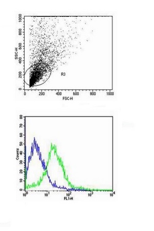
. Overlay histogram showing human PBMC cells stained with A01114-4 (Blue line). The cells were fixed with 4% paraformaldehyde and blocked with 10% normal goat serum. And then incubated with rabbit anti-TRAP/CD40L/CD40LG Antibody (A01114-4, 1microg/1x106 cells) for 30 min at 20°C. DyLight®488 conjugated goat anti-rabbit IgG (BA1127, 5-10microg/1x106 cells) was used as secondary antibody for 30 minutes at 20°C. Isotype control antibody (Green line) was rabbit IgG (1microg/1x106) used under the same conditions. Unlabelled sample without incubation with primary antibody and secondary antibody (Red line) was used as a blank control.")
Figure 1. Western blot analysis of TRAP/CD40L/CD40LG using anti-TRAP/CD40L/CD40LG antibody (A01114-4). Electrophoresis was performed on a 5-20% SDS-PAGE gel at 70V (Stacking gel) / 90V (Resolving gel) for 2-3 hours. The sample well of each lane was loaded with 30ug of sample under reducing conditions. Lane 1: human HL-60 whole cell lysates, Lane 2: human K562 whole cell lysates, Lane 3: human Raji whole cell lysates, Lane 4: human Jurkat whole cell lysates, Lane 5: rat RH35 whole cell lysates, Lane 6: mouse spleen tissue lysates, Lane 7: mouse ANA-1 whole cell lysates. After Electrophoresis, proteins were transferred to a Nitrocellulose membrane at 150mA for 50-90 minutes. Blocked the membrane with 5% Non-fat Milk/ TBS for 1.5 hour at RT. The membrane was incubated with rabbit anti-TRAP/CD40L/CD40LG antigen affinity purified polyclonal antibody (Catalog # A01114-4) at 0.5 microg/mL overnight at 4°C, then washed with TBS-0.1%Tween 3 times with 5 minutes each and probed with a goat anti-rabbit IgG-HRP secondary antibody at a dilution of 1:5000 for 1.5 hour at RT. The signal is developed using an Enhanced Chemiluminescent detection (ECL) kit (Catalog # EK1002) with Tanon 5200 system. A specific band was detected for TRAP/CD40L/CD40LG at approximately 36KD. The expected band size for TRAP/CD40L/CD40LG is at 36KD.
Anti-TRAP/CD40L/CD40LG Antibody Picoband(r)
A01114-4-DYLIGHT488
ApplicationsFlow Cytometry, Western Blot, ELISA
Product group Antibodies
ReactivityHuman, Mouse, Rat
TargetCD40LG
Overview
- SupplierBoster Bio
- Product NameAnti-TRAP/CD40L/CD40LG Antibody Picoband(r)
- Delivery Days Customer9
- ApplicationsFlow Cytometry, Western Blot, ELISA
- CertificationResearch Use Only
- ClonalityPolyclonal
- Concentration500 ug/ml
- ConjugateDyLight 488
- Gene ID959
- Target nameCD40LG
- Target descriptionCD40 ligand
- Target synonymsCD154, CD40L, HIGM1, IGM, IMD3, T-BAM, TNFSF5, TRAP, gp39, hCD40L, CD40 ligand, CD40 antigen ligand, CD40-L, T-B cell-activating molecule, T-cell antigen Gp39, TNF-related activation protein, tumor necrosis factor (ligand) superfamily member 5
- HostRabbit
- IsotypeIgG
- Protein IDP29965
- Protein NameCD40 ligand
- Scientific DescriptionBoster Bio Anti-TRAP/CD40L/CD40LG Antibody Picoband® catalog # A01114-4. Tested in ELISA, Flow Cytometry, WB applications. This antibody reacts with Human, Mouse, Rat. The brand Picoband indicates this is a premium antibody that guarantees superior quality, high affinity, and strong signals with minimal background in Western blot applications. Only our best-performing antibodies are designated as Picoband, ensuring unmatched performance.
- ReactivityHuman, Mouse, Rat
- Storage Instruction-20°C,2°C to 8°C
- UNSPSC12352203
Related products
Product group Antibodies
ApplicationsImmunoPrecipitation, Western Blot, ImmunoCytoChemistry, ImmunoHistoChemistry
ReactivityPorcine
TargetCD40LG
- SizePrice
Product group Antibodies
Anti-CD40LG AntibodyA39558
ApplicationsImmunoFluorescence, Western Blot
ReactivityHuman
- SizePrice
Product group Antibodies
ApplicationsFunctional Assay, Flow Cytometry, Western Blot, Neutralisation/Blocking
ReactivityCanine, Human
TargetCD40LG
- SizePrice
Product group Antibodies
ApplicationsFlow Cytometry, ImmunoPrecipitation
ReactivityHuman, Monkey
TargetCD40LG
- SizePrice
Product group Antibodies
References
CD40L Polyclonal AntibodyBS-1286R
ApplicationsFlow Cytometry, ImmunoFluorescence, Western Blot, ELISA, ImmunoCytoChemistry, ImmunoHistoChemistry, ImmunoHistoChemistry Frozen, ImmunoHistoChemistry Paraffin
ReactivityHuman, Rat
TargetCD40LG
- SizePrice
Product group Antibodies
ApplicationsFlow Cytometry
ReactivityHuman
TargetCD40LG
- SizePrice
Product group Antibodies

Anti-CD40L [hu5c8 (Ruplizumab)]Ab00447-1.1
ApplicationsFlow Cytometry, Western Blot, Neutralisation/Blocking
ReactivityHuman
TargetCD40LG
- SizePrice
Product group Antibodies
References
CD40L / CD154 antibody [5C8]GTX01484
ApplicationsFlow Cytometry
ReactivityHuman, Monkey, Primate
TargetCD40LG
- SizePrice
Product group Antibodies

CD40L Antibody (clone 5C8)LS-C770025
ApplicationsFlow Cytometry
ReactivityHuman, Monkey
TargetCD40LG
- SizePrice