

Antibodies
We offer one of the most comprehensive portfolios of antibodies. This includes monoclonal and polyclonal primary, secondary, conjugated, phospho-specific, functional, (isotype) controls, tagged and antibody pairs. In addition, we offer custom antibody services from several manufacturers.
The antibodies are generated in various hosts and react to antigens of different species like human, mouse, rat, rabbit or zebrafish. The antibodies are validated for multiple applications, including immunohistochemistry, western blot, immunoprecipitation, ELISA and flow cytometry, to ensure reliable performance for your research needs.
If you need a specific antibody and can’t find it in our webshop, please contact our technical support.
Discover what our customers say about us by reading their reviews.
![IHC-P analysis of human thyroid tissue using GTX47974 TSH receptor antibody [4C1].](https://www.genetex.com/upload/website/prouct_img/normal/GTX47974/GTX47974_5630_IHC-P_w_23060823_114.webp)
- SizePrice
![WB analysis of human brain tissue lysate using GTX48035 SSEA-1 antibody[MC-480].](https://www.genetex.com/upload/website/prouct_img/normal/GTX48035/GTX48035_1535_WB_w_23060823_356.webp)
- SizePrice
- SizePrice
- SizePrice
- SizePrice
- SizePrice
- SizePrice
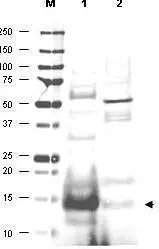
![Western blot analysis of GeneTex's IgY fraction of Chicken-anti-Human MIF polyclonal antibody shows the detection of 100 μg of recombinant MIF present in a lysate. Similar detection of MIF will occur when human serum is analyzed. In lane 1 no reaction is observed in the control whereas lane 2 shows a single band at 12.3 kDa. A 4-20% gradient gel was used to separate the proteins by SDS-PAGE. The protein was transferred to nitro-cellulose using standard methods. After blocking the membrane was probed with the primary antibody for 1 h at room temperature followed by washes and reaction with a 1:5,000 dilution of IRDye800 conjugated Gt-a-Chicken Rabbit IgG [H&L] for 1 h at room temperature. LICOR's OdysseyR Infrared Imaging System was used to scan and process the image. Other detection systems will yield similar results.](https://www.genetex.com/upload/website/prouct_img/normal/GTX48478/GTX48478_20160330_WB_w_23060823_846.webp)
- SizePrice
- SizePrice
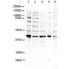
![Western blot analysis is shown using GeneTex's Affinity Purified anti-p28 ING5 antibody to detect over expressed Human ING5 present in cell extracts. This western blot shows reactivity with purified recombinant human ING5 protein. Comparison to a molecular weight marker (not shown) indicates a single band of ~36 kDa corresponding to the expected molecular weight for the recombinant protein. Approximately 10 μg of lysate was separated on a 4-20% Tris-Glycine gel by SDS-PAGE and transferred onto nitrocellulose. After blocking the membrane was probed with the primary antibody diluted to 1:1,500.Incubation was overnight at 4o C followed by washes and reaction with a 1:20,000 dilution of IRDye?800 conjugated Rb-a-Goat IgG [H&L] MXHu for 45 min at room temperature. IRDye?800 fluorescence image was captured using the OdysseyR Infrared Imaging System developed by LI-COR. IRDye is a trademark of LI-COR, Inc. Other detection systems will yield similar results.](https://www.genetex.com/upload/website/prouct_img/normal/GTX48484/GTX48484_20160330_WB_1_w_23060823_253.webp)
- SizePrice
- SizePrice
- SizePrice
Didn't find what you were looking for?
Search through our product groups to find the right product
Back to overview