


Antibodies
We offer one of the most comprehensive portfolios of antibodies. This includes monoclonal and polyclonal primary, secondary, conjugated, phospho-specific, functional, (isotype) controls, tagged and antibody pairs. In addition, we offer custom antibody services from several manufacturers.
The antibodies are generated in various hosts and react to antigens of different species like human, mouse, rat, rabbit or zebrafish. The antibodies are validated for multiple applications, including immunohistochemistry, western blot, immunoprecipitation, ELISA and flow cytometry, to ensure reliable performance for your research needs.
If you need a specific antibody and can’t find it in our webshop, please contact our technical support.
Discover what our customers say about us by reading their reviews.
- SizePrice
- SizePrice
- SizePrice
- SizePrice
- SizePrice
- SizePrice
- SizePrice
- SizePrice
- SizePrice

- SizePrice
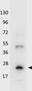
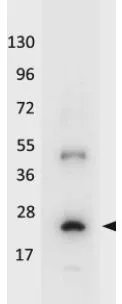
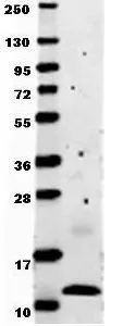
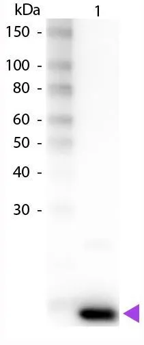
![Western blot of mammalian whole cell extract transfected with HA epitope tagged human ATF3. GeneTex's Affinity purified anti-ATF3 detects a band ~31 kDa corresponding to recombinant human ATF3. Immunostaining using GeneTex's anti-HA epitope tag antibody confirms the composition of the recombinant band (not shown). The protein was transferred to nitrocellulose in 30 minutes using standard methods. After blocking with 5% goat serum and 0.5% non-fat milk in PBS, the membrane was probed with the primary antibody diluted 1:200 in 0.2X blocking buffer in PBS overnight at 4oC. Reaction was followed by washes and reaction with a 1:5000 dilution of IRDye?800 conjugated Gt-a-Rabbit IgG [H&L] for 30 min at room temperature. LICOR's OdysseyR Infrared Imaging System was used to scan and process the image. Other detection systems will yield similar results.](https://www.genetex.com/upload/website/prouct_img/normal/GTX48669/GTX48669_20160330_WB_w_23060823_381.webp)
- SizePrice
- SizePrice
Didn't find what you were looking for?
Search through our product groups to find the right product
Back to overview