

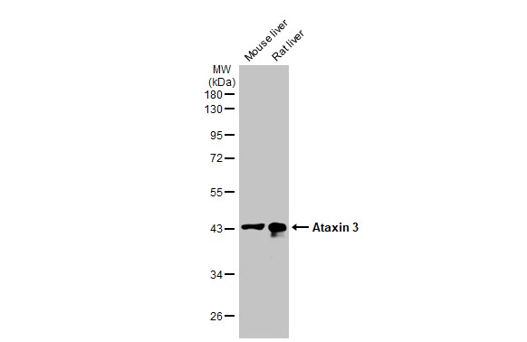
Various tissue extracts (50 μg) were separated by 10% SDS-PAGE, and the membrane was blotted with Ataxin 3 antibody [HL1909] (GTX637658) diluted at 1:1000. The HRP-conjugated anti-rabbit IgG antibody (GTX213110-01) was used to detect the primary antibody, and the signal was developed with Trident ECL plus-Enhanced.
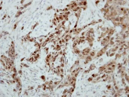
![Ataxin 3 antibody [HL1909] detects Ataxin 3 protein at nucleus by immunohistochemical analysis. Sample: Paraffin-embedded human lung cancer. Ataxin 3 stained by Ataxin 3 antibody [HL1909] (GTX637658) diluted at 1:100. Antigen Retrieval: Citrate buffer, pH 6.0, 15 min](https://www.genetex.com/upload/website/prouct_img/normal/GTX637658/GTX637658_T-44837_20221223_IHC-P_22122722_218.webp "Ataxin 3 antibody [HL1909] detects Ataxin 3 protein at nucleus by immunohistochemical analysis. Sample: Paraffin-embedded human lung cancer. Ataxin 3 stained by Ataxin 3 antibody [HL1909] (GTX637658) diluted at 1:100. Antigen Retrieval: Citrate buffer, pH 6.0, 15 min")
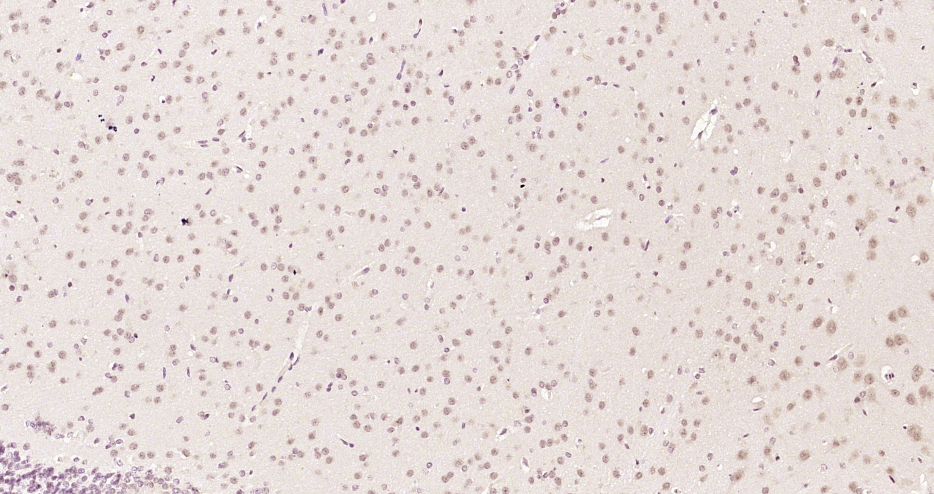
![Ataxin 3 antibody [HL1909] detects Ataxin 3 protein at nucleus by immunohistochemical analysis. Sample: Paraffin-embedded rat brain. Ataxin 3 stained by Ataxin 3 antibody [HL1909] (GTX637658) diluted at 1:100. Antigen Retrieval: Citrate buffer, pH 6.0, 15 min](https://www.genetex.com/upload/website/prouct_img/normal/GTX637658/GTX637658_T-44837_20221223_IHC-P_R_22122722_521.webp "Ataxin 3 antibody [HL1909] detects Ataxin 3 protein at nucleus by immunohistochemical analysis. Sample: Paraffin-embedded rat brain. Ataxin 3 stained by Ataxin 3 antibody [HL1909] (GTX637658) diluted at 1:100. Antigen Retrieval: Citrate buffer, pH 6.0, 15 min")
![Ataxin 3 antibody [HL1909] detects Ataxin 3 protein at nucleus by immunohistochemical analysis. Sample: Paraffin-embedded mouse stomach. Ataxin 3 stained by Ataxin 3 antibody [HL1909] (GTX637658) diluted at 1:100. Antigen Retrieval: Citrate buffer, pH 6.0, 15 min](https://www.genetex.com/upload/website/prouct_img/normal/GTX637658/GTX637658_T-44837_20221223_IHC-P_M_22122722_497.webp "Ataxin 3 antibody [HL1909] detects Ataxin 3 protein at nucleus by immunohistochemical analysis. Sample: Paraffin-embedded mouse stomach. Ataxin 3 stained by Ataxin 3 antibody [HL1909] (GTX637658) diluted at 1:100. Antigen Retrieval: Citrate buffer, pH 6.0, 15 min")
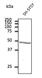
![Various whole cell extracts (30 μg) were separated by 10% SDS-PAGE, and the membrane was blotted with Ataxin 3 antibody [HL1909] (GTX637658) diluted at 1:1000. The HRP-conjugated anti-rabbit IgG antibody (GTX213110-01) was used to detect the primary antibody.](https://www.genetex.com/upload/website/prouct_img/normal/GTX637658/GTX637658_44900_20221223_WB_22122722_462.webp "Various whole cell extracts (30 μg) were separated by 10% SDS-PAGE, and the membrane was blotted with Ataxin 3 antibody [HL1909] (GTX637658) diluted at 1:1000. The HRP-conjugated anti-rabbit IgG antibody (GTX213110-01) was used to detect the primary antibody.")
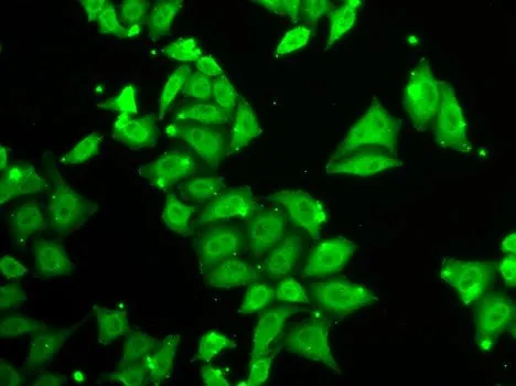
![Ataxin 3 antibody [HL1909] detects Ataxin 3 protein by immunofluorescent analysis. Sample: MCF-7 cells were fixed in 4% paraformaldehyde at RT for 15 min. Green: Ataxin 3 stained by Ataxin 3 antibody [HL1909] (GTX637658) diluted at 1:500. Red: alpha Tubulin, a cytoskeleton marker, stained by alpha Tubulin antibody [GT114] (GTX628802) diluted at 1:1000. Blue: Fluoroshield with DAPI (GTX30920). Scale bar= 10μm.](https://www.genetex.com/upload/website/prouct_img/normal/GTX637658/GTX637658_44900_20221230_ICC_IF_22122901_869.webp "Ataxin 3 antibody [HL1909] detects Ataxin 3 protein by immunofluorescent analysis. Sample: MCF-7 cells were fixed in 4% paraformaldehyde at RT for 15 min. Green: Ataxin 3 stained by Ataxin 3 antibody [HL1909] (GTX637658) diluted at 1:500. Red: alpha Tubulin, a cytoskeleton marker, stained by alpha Tubulin antibody [GT114] (GTX628802) diluted at 1:1000. Blue: Fluoroshield with DAPI (GTX30920). Scale bar= 10μm.")
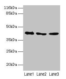
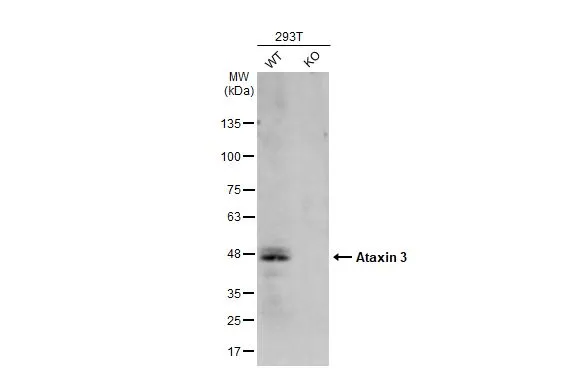
![Non-transfected (–) and transfected (+) 293T whole cell extracts (30 μg) were separated by 10% SDS-PAGE, and the membrane was blotted with Ataxin 3 antibody [HL1909] (GTX637658) diluted at 1:500. The HRP-conjugated anti-rabbit IgG antibody (GTX213110-01) was used to detect the primary antibody.](https://www.genetex.com/upload/website/prouct_img/normal/GTX637658/GTX637658_44900_20230210_WB_shRNA_watermark_23021401_927.webp "Non-transfected (–) and transfected (+) 293T whole cell extracts (30 μg) were separated by 10% SDS-PAGE, and the membrane was blotted with Ataxin 3 antibody [HL1909] (GTX637658) diluted at 1:500. The HRP-conjugated anti-rabbit IgG antibody (GTX213110-01) was used to detect the primary antibody.")
Various tissue extracts (50 μg) were separated by 10% SDS-PAGE, and the membrane was blotted with Ataxin 3 antibody [HL1909] (GTX637658) diluted at 1:1000. The HRP-conjugated anti-rabbit IgG antibody (GTX213110-01) was used to detect the primary antibody, and the signal was developed with Trident ECL plus-Enhanced.
Ataxin 3 antibody [HL1909]
GTX637658
ApplicationsImmunoFluorescence, Western Blot, ImmunoCytoChemistry, ImmunoHistoChemistry, ImmunoHistoChemistry Paraffin
Product group Antibodies
ReactivityHuman, Mouse, Rat
TargetATXN3
Overview
- SupplierGeneTex
- Product NameAtaxin 3 antibody [HL1909]
- Delivery Days Customer9
- Application Supplier NoteWB: 1:500-1:3000. *Optimal dilutions/concentrations should be determined by the researcher.Not tested in other applications.
- ApplicationsImmunoFluorescence, Western Blot, ImmunoCytoChemistry, ImmunoHistoChemistry, ImmunoHistoChemistry Paraffin
- CertificationResearch Use Only
- ClonalityMonoclonal
- Clone IDHL1909
- Concentration1 mg/ml
- ConjugateUnconjugated
- Gene ID4287
- Target nameATXN3
- Target descriptionataxin 3
- Target synonymsAT3, ATX3, JOS, MJD, MJD1, SCA3, ataxin-3, Machado-Joseph disease protein 1, josephin, olivopontocerebellar ataxia 3, spinocerebellar ataxia type 3 protein
- HostRabbit
- IsotypeIgG
- Protein IDP54252
- Protein NameAtaxin-3
- Scientific DescriptionMachado-Joseph disease, also known as spinocerebellar ataxia-3, is an autosomal dominant neurologic disorder. The protein encoded by this gene contains (CAG)n repeats in the coding region, and the expansion of these repeats from the normal 12-44 to 52-86 is one cause of Machado-Joseph disease. There is a negative correlation between the age of onset and CAG repeat numbers. Alternatively spliced transcript variants encoding different isoforms have been described for this gene. [provided by RefSeq, Jul 2016]
- ReactivityHuman, Mouse, Rat
- Storage Instruction-20°C or -80°C,2°C to 8°C
- UNSPSC41116161
Datasheet
Related products
Product group Antibodies
Anti-ATXN3 AntibodyA121608
ApplicationsWestern Blot
ReactivityCanine, Human, Monkey, Mouse, Rat
- SizePrice
Product group Antibodies
Anti-ATXN3 Antibody144-01243
ApplicationsImmunoFluorescence, Western Blot
ReactivityHuman
TargetATXN3
- SizePrice
Product group Antibodies
ATXN3 AntibodyCSB-PA002443LA01HU
ApplicationsWestern Blot, ELISA, ImmunoHistoChemistry
ReactivityHuman, Mouse
TargetATXN3
- SizePrice
Product group Antibodies
Atxn3 Polyclonal AntibodyCAC08255
ApplicationsWestern Blot, ELISA, ImmunoHistoChemistry
ReactivityMouse
TargetATXN3
- SizePrice
Product group Antibodies
Ataxin 3 Polyclonal AntibodyBS-17208R
ApplicationsImmunoFluorescence, Western Blot, ELISA, ImmunoCytoChemistry, ImmunoHistoChemistry, ImmunoHistoChemistry Frozen, ImmunoHistoChemistry Paraffin
ReactivityBovine, Canine, Equine, Human, Mouse, Porcine, Rat, Sheep
TargetATXN3
- SizePrice
Product group Antibodies
Ataxin 3 antibodyGTX30079
ApplicationsImmunoFluorescence, Western Blot, ImmunoCytoChemistry, ImmunoHistoChemistry, ImmunoHistoChemistry Paraffin
ReactivityHuman, Mouse, Rat
TargetATXN3
- SizePrice
Product group Antibodies
Ataxin 3 antibodyGTX101343
ApplicationsImmunoFluorescence, ImmunoPrecipitation, Western Blot, ImmunoCytoChemistry
ReactivityHuman
TargetATXN3
- SizePrice

Product group Antibodies
Anti-ATXN3 AntibodyHPA069338
ApplicationsImmunoCytoChemistry
ReactivityHuman
TargetATXN3
- SizePrice
Product group Antibodies
Ataxin 3 antibodyGTX115032
ApplicationsImmunoPrecipitation, Western Blot, ImmunoHistoChemistry, ImmunoHistoChemistry Paraffin
ReactivityHuman, Mouse, Zebra Fish
TargetATXN3
- SizePrice