

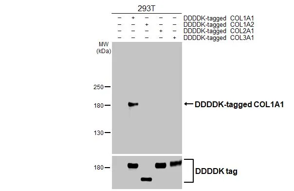
Non-transfected (–) and transfected (+) 293T whole cell extracts were separated by 5% SDS-PAGE, and the membrane was blotted with COL1A1 antibody [HL3040] (GTX640483) diluted at 1:5000. The HRP-conjugated anti-rabbit IgG antibody (GTX213110-01) was used to detect the primary antibody.
![COL1A1 antibody [HL3040] detects COL1A1 protein by immunofluorescent analysis. Sample: SK-N-SH cells were fixed in 4% paraformaldehyde at RT for 15 min. Green: COL1A1 stained by COL1A1 antibody [HL3040] (GTX640483) diluted at 1:500. Red: alpha Tubulin, a cytoskeleton marker, stained by alpha Tubulin antibody [GT114] (GTX628802) diluted at 1:1000. Blue: Fluoroshield with DAPI (GTX30920).](https://www.genetex.com/upload/website/prouct_img/normal/GTX640483/GTX640483_45551_20241213_ICC_IF_24123021_520.webp "COL1A1 antibody [HL3040] detects COL1A1 protein by immunofluorescent analysis. Sample: SK-N-SH cells were fixed in 4% paraformaldehyde at RT for 15 min. Green: COL1A1 stained by COL1A1 antibody [HL3040] (GTX640483) diluted at 1:500. Red: alpha Tubulin, a cytoskeleton marker, stained by alpha Tubulin antibody [GT114] (GTX628802) diluted at 1:1000. Blue: Fluoroshield with DAPI (GTX30920).")
![Various whole cell extracts (30 μg) were separated by 5% SDS-PAGE, and the membrane was blotted with COL1A1 antibody [HL3040] (GTX640483) diluted at 1:1000. The HRP-conjugated anti-rabbit IgG antibody (GTX213110-01) was used to detect the primary antibody.](https://www.genetex.com/upload/website/prouct_img/normal/GTX640483/GTX640483_T-45439_20250103_WB_M_25010819_301.webp "Various whole cell extracts (30 μg) were separated by 5% SDS-PAGE, and the membrane was blotted with COL1A1 antibody [HL3040] (GTX640483) diluted at 1:1000. The HRP-conjugated anti-rabbit IgG antibody (GTX213110-01) was used to detect the primary antibody.")
![Various whole cell extracts (30 μg) were separated by 5% SDS-PAGE, and the membrane was blotted with COL1A1 antibody [HL3040] (GTX640483) diluted at 1:1000. The HRP-conjugated anti-rabbit IgG antibody (GTX213110-01) was used to detect the primary antibody. Corresponding RNA expression data for the same cell lines are based on Human Protein Atlas program.](https://www.genetex.com/upload/website/prouct_img/normal/GTX640483/GTX640483_T-45439_20250103_WB_TPM_watermark_25010819_360.webp "Various whole cell extracts (30 μg) were separated by 5% SDS-PAGE, and the membrane was blotted with COL1A1 antibody [HL3040] (GTX640483) diluted at 1:1000. The HRP-conjugated anti-rabbit IgG antibody (GTX213110-01) was used to detect the primary antibody. Corresponding RNA expression data for the same cell lines are based on Human Protein Atlas program.")
![Various whole cell extracts (30 μg) were separated by 5% SDS-PAGE, and the membrane was blotted with COL1A1 antibody [HL3040] (GTX640483) diluted at 1:1000. The HRP-conjugated anti-rabbit IgG antibody (GTX213110-01) was used to detect the primary antibody.](https://www.genetex.com/upload/website/prouct_img/normal/GTX640483/GTX640483_T-45439_20250103_WB_R_25010819_586.webp "Various whole cell extracts (30 μg) were separated by 5% SDS-PAGE, and the membrane was blotted with COL1A1 antibody [HL3040] (GTX640483) diluted at 1:1000. The HRP-conjugated anti-rabbit IgG antibody (GTX213110-01) was used to detect the primary antibody.")
Non-transfected (–) and transfected (+) 293T whole cell extracts were separated by 5% SDS-PAGE, and the membrane was blotted with COL1A1 antibody [HL3040] (GTX640483) diluted at 1:5000. The HRP-conjugated anti-rabbit IgG antibody (GTX213110-01) was used to detect the primary antibody.
COL1A1 antibody [HL3040]
GTX640483
ApplicationsImmunoFluorescence, Western Blot, ImmunoCytoChemistry
Product group Antibodies
ReactivityHuman, Mouse, Rat
TargetCOL1A1
Overview
- SupplierGeneTex
- Product NameCOL1A1 antibody [HL3040]
- Delivery Days Customer7
- Application Supplier NoteWB: 1:1000-1:10000. *Optimal dilutions/concentrations should be determined by the researcher.Not tested in other applications.
- ApplicationsImmunoFluorescence, Western Blot, ImmunoCytoChemistry
- CertificationResearch Use Only
- ClonalityMonoclonal
- Clone IDHL3040
- Concentration1 mg/ml
- ConjugateUnconjugated
- Gene ID1277
- Target nameCOL1A1
- Target descriptioncollagen type I alpha 1 chain
- Target synonymsCAFYD, EDSARTH1, EDSC, OI1, OI2, OI3, OI4, collagen alpha-1(I) chain, alpha-1 type I collagen, alpha1(I) procollagen, collagen Col1-ColIII-1, collagen Col1-ColIII-2, collagen alpha 1 chain type I, collagen alpha-1(I) chain preproprotein, collagen of skin, tendon and bone, alpha-1 chain, collagen, type I, alpha 1, pro-alpha-1 collagen type 1, type I proalpha 1, type I procollagen alpha 1 chain
- HostRabbit
- IsotypeIgG
- Protein IDP02452
- Protein NameCollagen alpha-1(I) chain
- Scientific DescriptionThis gene encodes the pro-alpha1 chains of type I collagen whose triple helix comprises two alpha1 chains and one alpha2 chain. Type I is a fibril-forming collagen found in most connective tissues and is abundant in bone, cornea, dermis and tendon. Mutations in this gene are associated with osteogenesis imperfecta types I-IV, Ehlers-Danlos syndrome type VIIA, Ehlers-Danlos syndrome Classical type, Caffey Disease and idiopathic osteoporosis. Reciprocal translocations between chromosomes 17 and 22, where this gene and the gene for platelet-derived growth factor beta are located, are associated with a particular type of skin tumor called dermatofibrosarcoma protuberans, resulting from unregulated expression of the growth factor. Two transcripts, resulting from the use of alternate polyadenylation signals, have been identified for this gene. [provided by R. Dalgleish, Feb 2008]
- ReactivityHuman, Mouse, Rat
- Storage Instruction-20°C or -80°C,2°C to 8°C
- UNSPSC41116161
Datasheet
Related products
Product group Antibodies

Collagen Type I, humanCO20111-0.1
ApplicationsImmunoFluorescence, Western Blot, ELISA, ImmunoHistoChemistry, ImmunoHistoChemistry Paraffin, RadioImmunoAssay
TargetCOL1A1
- SizePrice
Product group Antibodies
Anti-COL1A1 Antibody144-62810
ApplicationsImmunoFluorescence, ImmunoPrecipitation, Western Blot, ImmunoHistoChemistry
ReactivityHuman, Mouse, Rat
TargetCOL1A1
- SizePrice
Product group Antibodies
Anti-Collagen I AntibodyA95083
ApplicationsImmunoFluorescence, ELISA, ImmunoHistoChemistry
ReactivityHuman, Mouse, Rat
- SizePrice
Product group Antibodies

Anti-collagen type I [3P1-31], Human IgG1-Fc Fusion,AB04222-10.159
ApplicationsELISA
ReactivityHuman
TargetCOL1A1
- SizePrice
Product group Antibodies

COL1A1 / Collagen I Alpha 1 AntibodyLS-C831483
ApplicationsImmunoHistoChemistry
ReactivityHuman
TargetCOL1A1
- SizePrice

Product group Antibodies
Anti-COL1A1 AntibodyHPA008405
ApplicationsImmunoHistoChemistry
ReactivityHuman
TargetCOL1A1
- SizePrice
Product group Antibodies
COL1A1 Monoclonal AntibodyCSB-MA139659
ApplicationsELISA, ImmunoHistoChemistry
ReactivityHuman, Mouse, Rat
TargetCOL1A1
- SizePrice
Product group Antibodies
COL1A2/COL1A1 AntibodyPACO07262
ApplicationsWestern Blot, ELISA
ReactivityHuman, Mouse, Rat
TargetCOL1A1
- SizePrice
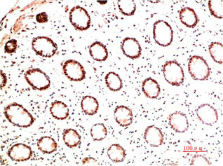
![Collagen I + Collagen II + Collagen III antibody [HL2048 + HL1907] detects secreted Collagen I + Collagen II + Collagen III protein by immunohistochemical analysis. Sample: Paraffin-embedded rat kidney. Collagen I + Collagen II + Collagen III stained by Collagen I + Collagen II + Collagen III antibody [HL2048 + HL1907] (GTX638633) diluted at 1:100. Antigen Retrieval: Citrate buffer, pH 6.0, 15 min](https://www.genetex.com/upload/website/prouct_img/normal/GTX638633/GTX638633_T-45061_20230616_IHC-P_R_23062718_561.webp)
Product group Antibodies
ApplicationsWestern Blot, ImmunoHistoChemistry, ImmunoHistoChemistry Paraffin
ReactivityHuman, Mouse, Rat
TargetCOL1A1
- SizePrice