

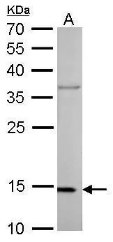
COX4 antibody detects COX4I1 protein by western blot analysis. A. 50 μg mouse heart lysate/extract 12% SDS-PAGE COX4 antibody (GTX628901) dilution: 1:1000 The HRP-conjugated anti-mouse IgG antibody (GTX213111-01) was used to detect the primary antibody.
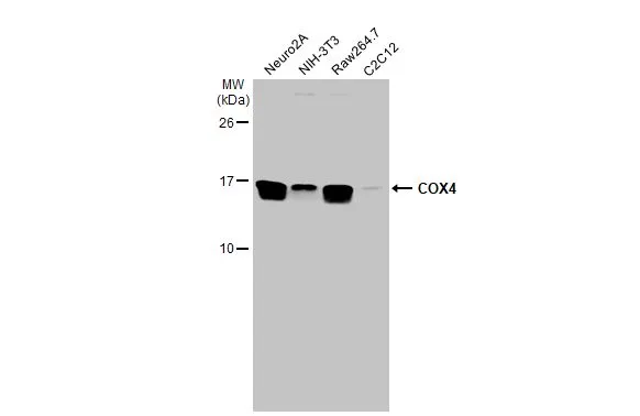
![Various whole cell extracts (30 μg) were separated by 15% SDS-PAGE, and the membrane was blotted with COX4 antibody [GT854] (GTX628901) diluted at 1:1000. The HRP-conjugated anti-mouse IgG antibody (GTX213111-01) was used to detect the primary antibody.](https://www.genetex.com/upload/website/prouct_img/normal/GTX628901/GTX628901_41253_20190809_WB_w_23061202_606.webp "Various whole cell extracts (30 μg) were separated by 15% SDS-PAGE, and the membrane was blotted with COX4 antibody [GT854] (GTX628901) diluted at 1:1000. The HRP-conjugated anti-mouse IgG antibody (GTX213111-01) was used to detect the primary antibody.")
 diluted at 1:1000.")
![COX4 antibody [GT854] detects COX4 protein at mitochondria by immunohistochemical analysis. Sample: Paraffin-embedded human colon cancer. COX4 stained by COX4 antibody [GT854] (GTX628901) diluted at 1:200. Antigen Retrieval: Tris-EDTA buffer, pH 9.0, 15 min](https://www.genetex.com/upload/website/prouct_img/normal/GTX628901/GTX628901_41253_20181025_IHC-P_1_w_23061202_110.webp "COX4 antibody [GT854] detects COX4 protein at mitochondria by immunohistochemical analysis. Sample: Paraffin-embedded human colon cancer. COX4 stained by COX4 antibody [GT854] (GTX628901) diluted at 1:200. Antigen Retrieval: Tris-EDTA buffer, pH 9.0, 15 min")
![COX4 antibody [GT854] detects COX4 protein at mitochondria by immunohistochemical analysis. Sample: Paraffin-embedded human ovarian cancer. COX4 stained by COX4 antibody [GT854] (GTX628901) diluted at 1:200. Antigen Retrieval: Tris-EDTA buffer, pH 9.0, 15 min](https://www.genetex.com/upload/website/prouct_img/normal/GTX628901/GTX628901_41253_20181025_IHC-P_2_w_23061202_264.webp "COX4 antibody [GT854] detects COX4 protein at mitochondria by immunohistochemical analysis. Sample: Paraffin-embedded human ovarian cancer. COX4 stained by COX4 antibody [GT854] (GTX628901) diluted at 1:200. Antigen Retrieval: Tris-EDTA buffer, pH 9.0, 15 min")
![COX4 antibody [GT854] detects COX4I1 protein by western blot analysis. A. 50 μg rat muscle lysate/extract 15% SDS-PAGE COX4 antibody [GT854] (GTX628901) dilution: 1:1000 The HRP-conjugated anti-mouse IgG antibody (GTX213111-01) was used to detect the primary antibody.](https://www.genetex.com/upload/website/prouct_img/normal/GTX628901/GTX628901_41253_WB_R_muscle_w_23061202_406.webp "COX4 antibody [GT854] detects COX4I1 protein by western blot analysis. A. 50 μg rat muscle lysate/extract 15% SDS-PAGE COX4 antibody [GT854] (GTX628901) dilution: 1:1000 The HRP-conjugated anti-mouse IgG antibody (GTX213111-01) was used to detect the primary antibody.")
![Non-transfected (–) and transfected (+) 293T whole cell extracts (30 μg) were separated by 15% SDS-PAGE, and the membrane was blotted with COX4 antibody [GT854] (GTX628901) diluted at 1:1500. The HRP-conjugated anti-mouse IgG antibody (GTX213111-01) was used to detect the primary antibody.](https://www.genetex.com/upload/website/prouct_img/normal/GTX628901/GTX628901_41253_20160721_WB_shRNA_watermark_w_23061202_222.webp "Non-transfected (–) and transfected (+) 293T whole cell extracts (30 μg) were separated by 15% SDS-PAGE, and the membrane was blotted with COX4 antibody [GT854] (GTX628901) diluted at 1:1500. The HRP-conjugated anti-mouse IgG antibody (GTX213111-01) was used to detect the primary antibody.")
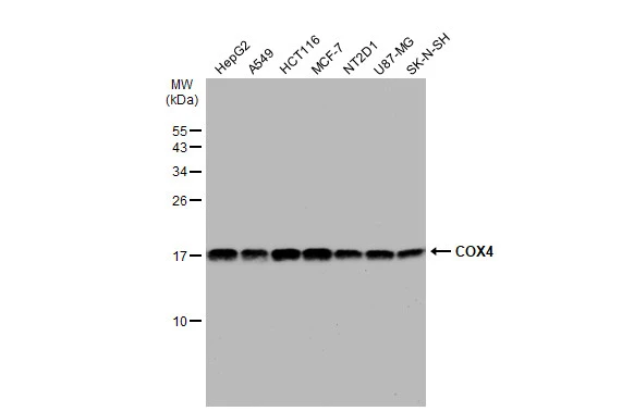
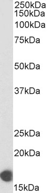
![Various tissue extracts (50 μg) were separated by 15% SDS-PAGE, and the membrane was blotted with COX4 antibody [GT854] (GTX628901) diluted at 1:1000. The HRP-conjugated anti-mouse IgG antibody (GTX213111-01) was used to detect the primary antibody.](https://www.genetex.com/upload/website/prouct_img/normal/GTX628901/GTX628901_41253_20191018_WB_M_tissue_w_23061202_396.webp "Various tissue extracts (50 μg) were separated by 15% SDS-PAGE, and the membrane was blotted with COX4 antibody [GT854] (GTX628901) diluted at 1:1000. The HRP-conjugated anti-mouse IgG antibody (GTX213111-01) was used to detect the primary antibody.")
COX4 antibody detects COX4I1 protein by western blot analysis. A. 50 μg mouse heart lysate/extract 12% SDS-PAGE COX4 antibody (GTX628901) dilution: 1:1000 The HRP-conjugated anti-mouse IgG antibody (GTX213111-01) was used to detect the primary antibody.
COX4 antibody [GT854]
GTX628901
ApplicationsImmunoFluorescence, Western Blot, ImmunoCytoChemistry, ImmunoHistoChemistry, ImmunoHistoChemistry Paraffin
Product group Antibodies
ReactivityHuman, Mouse, Rat
TargetCOX4I1
Overview
- SupplierGeneTex
- Product NameCOX4 antibody [GT854]
- Delivery Days Customer9
- Application Supplier NoteWB: 1:500-1:3000. ICC/IF: 1:100-1:1000. IHC-P: 1:100-1:1000. *Optimal dilutions/concentrations should be determined by the researcher.Not tested in other applications.
- ApplicationsImmunoFluorescence, Western Blot, ImmunoCytoChemistry, ImmunoHistoChemistry, ImmunoHistoChemistry Paraffin
- CertificationResearch Use Only
- ClonalityMonoclonal
- Clone IDGT854
- Concentration1 mg/ml
- ConjugateUnconjugated
- Gene ID1327
- Target nameCOX4I1
- Target descriptioncytochrome c oxidase subunit 4I1
- Target synonymsCOX IV-1, COX4, COX4-1, COXIV, COXIV-1, MC4DN16, cytochrome c oxidase subunit 4 isoform 1, mitochondrial, cytochrome c oxidase polypeptide IV, cytochrome c oxidase subunit IV
- HostMouse
- IsotypeIgG2a
- Protein IDP13073
- Protein NameCytochrome c oxidase subunit 4 isoform 1, mitochondrial
- Scientific DescriptionCytochrome c oxidase (COX) is the terminal enzyme of the mitochondrial respiratory chain. It is a multi-subunit enzyme complex that couples the transfer of electrons from cytochrome c to molecular oxygen and contributes to a proton electrochemical gradient across the inner mitochondrial membrane. The complex consists of 13 mitochondrial- and nuclear-encoded subunits. The mitochondrially-encoded subunits perform the electron transfer and proton pumping activities. The functions of the nuclear-encoded subunits are unknown but they may play a role in the regulation and assembly of the complex. This gene encodes the nuclear-encoded subunit IV isoform 1 of the human mitochondrial respiratory chain enzyme. It is located at the 3 of the NOC4 (neighbor of COX4) gene in a head-to-head orientation, and shares a promoter with it. [provided by RefSeq]
- ReactivityHuman, Mouse, Rat
- Storage Instruction-20°C or -80°C,2°C to 8°C
- UNSPSC12352203
References
- Perić I, Costina V, Findeisen P, et al. Tianeptine Enhances Energy-related Processes in the Hippocampal Non-synaptic Mitochondria in a Rat Model of Depression. Neuroscience. 2020,451:111-125. doi: 10.1016/j.neuroscience.2020.09.061Read this paper
- Zhao J, Fu X, Chen H, et al. G3BP1 interacts with YWHAZ to regulate chemoresistance and predict adjuvant chemotherapy benefit in gastric cancer. Br J Cancer. 2021,124(2):425-436. doi: 10.1038/s41416-020-01067-1Read this paper
- Dongil P, Pérez-García A, Hurtado-Carneiro V, et al. PAS kinase deficiency reduces aging effects in mice. Aging (Albany NY). 2020,12(3):2275-2301. doi: 10.18632/aging.102745Read this paper
Datasheet
Related products
Product group Antibodies
References
COX4 antibodyGTX101499
ApplicationsImmunoFluorescence, ImmunoPrecipitation, Western Blot, ImmunoCytoChemistry, ImmunoHistoChemistry, ImmunoHistoChemistry Paraffin
ReactivityHuman, Mouse
TargetCOX4I1
- SizePrice
Product group Antibodies
References
COX4 antibodyGTX114330
ApplicationsImmunoFluorescence, Western Blot, ImmunoCytoChemistry, ImmunoHistoChemistry, ImmunoHistoChemistry Frozen, ImmunoHistoChemistry Paraffin
ReactivityHuman, Mouse, Rat
TargetCOX4I1
- SizePrice
![Various whole cell extracts (30 μg) were separated by 15% SDS-PAGE, and the membrane was blotted with COX4 antibody [GT6310] (GTX628886) diluted at 1:1000. The HRP-conjugated anti-mouse IgG antibody (GTX213111-01) was used to detect the primary antibody.](https://www.genetex.com/upload/website/prouct_img/normal/GTX628886/GTX628886_44503_20211112_WB_22110201_302.webp)
Product group Antibodies
References
COX4 antibody [GT6310]GTX628886
ApplicationsImmunoFluorescence, Western Blot, ImmunoCytoChemistry, ImmunoHistoChemistry, ImmunoHistoChemistry Paraffin
ReactivityHuman, Rat
TargetCOX4I1
- SizePrice
![COX4 antibody [HL2264] detects COX4 protein by immunohistochemical analysis. Sample: Paraffin-embedded mouse tissues. COX4 stained by COX4 antibody [HL2264] (GTX638316) diluted at 1:100. Antigen Retrieval: Citrate buffer, pH 6.0, 15 min](https://www.genetex.com/upload/website/prouct_img/normal/GTX638316/GTX638316_T-44970_20230324_IHC-P_multiple_M_23032819_438.webp)
Product group Antibodies
COX4 antibody [HL2264]GTX638316
ApplicationsImmunoFluorescence, Western Blot, ImmunoCytoChemistry, ImmunoHistoChemistry, ImmunoHistoChemistry Paraffin
ReactivityHuman, Mouse, Rat, Zebra Fish
TargetCOX4I1
- SizePrice
![WB analysis of HEK293 (1), A549 (2) and PC12 (3) cell lysate using GTX82782 COX4 antibody [6B3].](https://www.genetex.com/upload/website/prouct_img/normal/GTX82782/GTX82782_20170912_WB_w_23061322_969.webp)
Product group Antibodies
COX4 antibody [6B3]GTX82782
ApplicationsFlow Cytometry, ImmunoFluorescence, Western Blot, ELISA, ImmunoCytoChemistry
ReactivityHuman, Monkey, Mouse, Rat
TargetCOX4I1
- SizePrice
Product group Antibodies
Cox4I1 Polyclonal AntibodyCAC07075
ApplicationsWestern Blot, ELISA, ImmunoHistoChemistry
ReactivityRat, Zebra Fish
TargetCOX4I1
- SizePrice
Product group Antibodies
References
COX4I1 Polyclonal AntibodyBS-10257R
ApplicationsFlow Cytometry, ImmunoFluorescence, Western Blot, ELISA, ImmunoCytoChemistry, ImmunoHistoChemistry, ImmunoHistoChemistry Frozen, ImmunoHistoChemistry Paraffin
ReactivityBovine, Canine, Equine, Human, Mouse, Porcine, Rat
TargetCOX4I1
- SizePrice
Product group Antibodies
ApplicationsWestern Blot, ELISA
ReactivityHuman
- SizePrice