

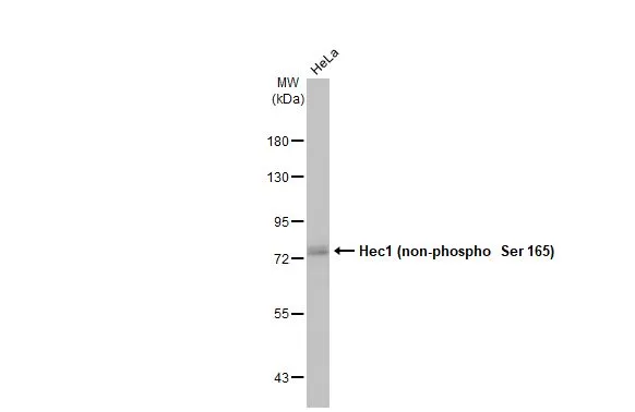
Whole cell extract (30 μg) was separated by 7.5% SDS-PAGE, and the membrane was blotted with Hec1 (non-phospho Ser 165) antibody (GTX70012) diluted at 1:1000. The HRP-conjugated anti-rabbit IgG antibody (GTX213110-01) was used to detect the primary antibody.
Whole cell extract (30 μg) was separated by 7.5% SDS-PAGE, and the membrane was blotted with Hec1 (non-phospho Ser 165) antibody (GTX70012) diluted at 1:1000. The HRP-conjugated anti-rabbit IgG antibody (GTX213110-01) was used to detect the primary antibody.
Hec1 (non-phospho Ser 165) antibody
GTX70012
ApplicationsImmunoFluorescence, Western Blot, ELISA, ImmunoCytoChemistry
Product group Antibodies
ReactivityHuman, Mouse, Rat
TargetNDC80
Overview
- SupplierGeneTex
- Product NameHec1 (non-phospho Ser 165) antibody
- Delivery Days Customer9
- Application Supplier NoteRecommended Starting Dilutions:For WB: Use at a dilution of 1:600For ICC/IF: Use at a dilution of 1:300Not yet tested in other applications. Optimal dilutions should be determined experimentally by the researcher.
- ApplicationsImmunoFluorescence, Western Blot, ELISA, ImmunoCytoChemistry
- CertificationResearch Use Only
- ClonalityPolyclonal
- Concentration0.91 mg/ml
- ConjugateUnconjugated
- Gene ID10403
- Target nameNDC80
- Target descriptionNDC80 kinetochore complex component
- Target synonymsHEC, HEC1, HsHec1, KNTC2, TID3, hsNDC80, kinetochore protein NDC80 homolog, NDC80 homolog, kinetochore complex component, NDC80 kinetochore complex component homolog, highly expressed in cancer protein, highly expressed in cancer, rich in leucine heptad repeats, kinetochore associated 2, kinetochore protein Hec1, kinetochore-associated protein 2, retinoblastoma-associated protein HEC
- HostRabbit
- IsotypeIgG
- Protein IDO14777
- Protein NameKinetochore protein NDC80 homolog
- Scientific DescriptionThis gene encodes a component of the NDC80 kinetochore complex. The encoded protein consists of an N-terminal microtubule binding domain and a C-terminal coiled-coiled domain that interacts with other components of the complex. This protein functions to organize and stabilize microtubule-kinetochore interactions and is required for proper chromosome segregation. [provided by RefSeq, Oct 2011]
- ReactivityHuman, Mouse, Rat
- Storage Instruction-20°C or -80°C,2°C to 8°C
- UNSPSC12352203
Datasheet
Related products
Product group Antibodies

Anti-Kinetochore protein NDC80 homolog [Hec1 (9G3)]AB03500-1.1-BT
ApplicationsImmunoFluorescence, ImmunoPrecipitation, Western Blot
ReactivityHuman
TargetNDC80
- SizePrice
Product group Antibodies
Anti-NDC80 Antibody144-05411
ApplicationsWestern Blot
ReactivityHuman, Mouse
TargetNDC80
- SizePrice

Product group Antibodies
Anti-HEC1/HEC/NDC80 Antibody Picoband(r)A01731-2-CARRIER-FREE
ApplicationsFlow Cytometry, ImmunoFluorescence, Western Blot, ELISA, ImmunoCytoChemistry
ReactivityHuman, Mouse, Rat
TargetNDC80
- SizePrice
Product group Antibodies
References
Hec1 antibodyGTX110735
ApplicationsImmunoPrecipitation, Western Blot
ReactivityHuman
TargetNDC80
- SizePrice
![WB analysis of (1) Jurkat, (2) HeLa, (3) NCCIT and (4) human spleen lysates (10 μg per lane) using HEC1 antibody [EPR5342] at a dilution of 1:10,000.](https://www.genetex.com/upload/website/prouct_img/normal/GTX63082/GTX63082_WB_w_23061202_241.webp)
Product group Antibodies
Hec1 antibody [EPR5342]GTX63082
ApplicationsImmunoPrecipitation, Western Blot
ReactivityHuman, Mouse, Rat
TargetNDC80
- SizePrice
Product group Antibodies
ApplicationsImmunoFluorescence, Western Blot, ELISA, ImmunoCytoChemistry
ReactivityHuman
TargetNDC80
- SizePrice

Product group Antibodies
ApplicationsImmunoFluorescence, Western Blot, ELISA, ImmunoCytoChemistry
ReactivityHuman
TargetNDC80
- SizePrice
Product group Antibodies
Hec1 antibodyGTX70016
ApplicationsImmunoFluorescence, ImmunoPrecipitation, Western Blot, ELISA, ImmunoCytoChemistry
ReactivityHuman, Mouse, Rat
TargetNDC80
- SizePrice

Product group Antibodies
References
Hec1 (phospho Ser 55) antibodyGTX70017
ApplicationsWestern Blot
ReactivityHuman, Mouse
TargetNDC80
- SizePrice
![Hec1 antibody [9G3.23] detects Hec1 protein at kinetochore by immunofluorescent analysis. Sample: HeLa cells were fixed in 4% paraformaldehyde at RT for 15 min. Green: Hec1 stained by Hec1 antibody [9G3.23] (GTX70268) diluted at 1:500. Red: beta Tubulin, a cytoskeleton marker, stained by beta Tubulin antibody (GTX101279) diluted at 1:1000. Blue: Fluoroshield with DAPI (GTX30920).](https://www.genetex.com/upload/website/prouct_img/normal/GTX70268/GTX70268_44482_20230119_ICC_IF_23021401_595.webp)
Product group Antibodies
References
Hec1 antibody [9G3.23]GTX70268
ApplicationsFlow Cytometry, ImmunoFluorescence, ImmunoPrecipitation, Western Blot, ImmunoCytoChemistry, ImmunoHistoChemistry, ImmunoHistoChemistry Frozen, Other Application
ReactivityHamster, Human, Mammals, Mouse
TargetNDC80
- SizePrice