

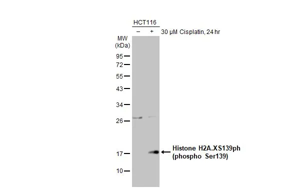
Untreated (–) and treated (+) HCT116 whole cell extracts (30 μg) were separated by 12% SDS-PAGE, and the membrane was blotted with Histone H2A.XS139ph (phospho Ser139) antibody [GT2311] (GTX628789) diluted at 1:1000. The HRP-conjugated anti-mouse IgG antibody (GTX213111-01) was used to detect the primary antibody, and the signal was developed with Trident ECL plus-Enhanced.
![Histone H2A.X (phospho S139) antibody [GT2311] detects H2AFX protein by western blot analysis. A. 30 μg NIH-3T3 whole cell lysate/extract (untreated) B. 30 μg NIH-3T3 whole cell lysate/extract (30μM cisplatin treatment for 24hr) 15% SDS-PAGE Histone H2A.X (phospho S139) antibody [GT2311] (GTX628789) dilution: 1:1000 The HRP-conjugated anti-mouse IgG antibody (GTX213111-01) was used to detect the primary antibody.](https://www.genetex.com/upload/website/prouct_img/normal/GTX628789/GTX628789_41190_WB_M_cisplatin_w_23061202_867.webp "Histone H2A.X (phospho S139) antibody [GT2311] detects H2AFX protein by western blot analysis. A. 30 μg NIH-3T3 whole cell lysate/extract (untreated) B. 30 μg NIH-3T3 whole cell lysate/extract (30μM cisplatin treatment for 24hr) 15% SDS-PAGE Histone H2A.X (phospho S139) antibody [GT2311] (GTX628789) dilution: 1:1000 The HRP-conjugated anti-mouse IgG antibody (GTX213111-01) was used to detect the primary antibody.")
 antibody immunoprecipitates histone H2A.X (phospho S139) protein in IP experiments. IP Sample: 500 μg HCT116 with CPT 30 μM treatment 24 hr whole cell lysate/extract A. 30 μg HCT116 whole with CPT 30 uM treatment cell lysate/extract B. Control with 2 μg of preimmune mouse IgG C. Immunoprecipitation of histone H2A.X (phospho S139) protein by 2 μg histone H2A.X (phospho S139) antibody (GTX628789) 15% SDS-PAGE The immunoprecipitated histone H2A.X (phospho S139) protein was detected by Human histone H2A.X (phospho S139) antibody (GTX628789) diluted at 1:1000. EasyBlot anti-mouse IgG (GTX221667-01) was used as a secondary reagent.")
![Histone H2A.XS139ph (phospho Ser139) antibody [GT2311] detects Histone H2A.XS139ph (phospho Ser139) protein at nucleus on mouse testis by immunohistochemical analysis. Sample: Paraffin-embedded mouse testis. Histone H2A.XS139ph (phospho Ser139) antibody [GT2311] (GTX628789) dilution: 1:500.
Antigen Retrieval: Trilogy? (EDTA based, pH 8.0) buffer, 15min](https://www.genetex.com/upload/website/prouct_img/normal/GTX628789/GTX628789_41190_IHC_M_w_23061202_485.webp "Histone H2A.XS139ph (phospho Ser139) antibody [GT2311] detects Histone H2A.XS139ph (phospho Ser139) protein at nucleus on mouse testis by immunohistochemical analysis. Sample: Paraffin-embedded mouse testis. Histone H2A.XS139ph (phospho Ser139) antibody [GT2311] (GTX628789) dilution: 1:500.
Antigen Retrieval: Trilogy? (EDTA based, pH 8.0) buffer, 15min")
![Histone H2A.X (phospho Ser139) antibody detects H2AFX protein at nuclear by confocal immunofluorescent analysis. Sample: 10μM Cisplatin treated (right) or untreated (left) HeLa cells were fixed in 4% paraformaldehyde for 15 min. Red: H2A.X protein stained by Histone H2A.X (phospho Ser139) antibody (GTX628789) diluted at 1:500. Green: alpha Tubulin antibody (GTX102078) diluted at 1:1000. Blue: Hoechst 33342 staining. [Images captured by Olympus FV1000 Confocal Laser Scanning Microscope]](https://www.genetex.com/upload/website/prouct_img/normal/GTX628789/GTX628789_41190_CT_IFA_w_23061202_917.webp "Histone H2A.X (phospho Ser139) antibody detects H2AFX protein at nuclear by confocal immunofluorescent analysis. Sample: 10μM Cisplatin treated (right) or untreated (left) HeLa cells were fixed in 4% paraformaldehyde for 15 min. Red: H2A.X protein stained by Histone H2A.X (phospho Ser139) antibody (GTX628789) diluted at 1:500. Green: alpha Tubulin antibody (GTX102078) diluted at 1:1000. Blue: Hoechst 33342 staining. [Images captured by Olympus FV1000 Confocal Laser Scanning Microscope]")
![Histone H2A.X (phospho S139) antibody [GT2311] detects Histone H2A.X (phospho S139) [GT2311] protein by western blot analysis. Un-treated (-) and treated (+, 30 μM Cisplatin treatment for 24 hrs) PC-12 whole cell extracts (30 μg) were separated by 15% SDS-PAGE, and the membrane was blotted with Histone H2A.X (phospho S139) antibody [GT2311] (GTX628789) diluted by 1:500. The HRP-conjugated anti-mouse IgG antibody (GTX213111-01) was used to detect the primary antibody.](https://www.genetex.com/upload/website/prouct_img/normal/GTX628789/GTX628789_41190_WB_Cisplatin_2_w_23061202_509.webp "Histone H2A.X (phospho S139) antibody [GT2311] detects Histone H2A.X (phospho S139) [GT2311] protein by western blot analysis. Un-treated (-) and treated (+, 30 μM Cisplatin treatment for 24 hrs) PC-12 whole cell extracts (30 μg) were separated by 15% SDS-PAGE, and the membrane was blotted with Histone H2A.X (phospho S139) antibody [GT2311] (GTX628789) diluted by 1:500. The HRP-conjugated anti-mouse IgG antibody (GTX213111-01) was used to detect the primary antibody.")
 or untreated (left) HeLa cells were fixed in 4% paraformaldehyde for 15 min. Green: H2AFX protein stained by Histone H2A.Xantibody (GTX628789) diluted at 1:500. Blue: Hoechst 33342 staining.")
 antibody detects Histone H2A.XS139ph (phospho Ser139) protein by western blot analysis. Un-treated (-) and treated (+, 50 J/m2 UV treatment) U2OS whole cell extracts (16 μg) were separated by 12%-15% SDS-PAGE, and the membrane was blotted with Histone H2A.XS139ph (phospho Ser139) antibody (GTX628789) diluted at 1:1000. The HRP-conjugated anti-mouse IgG antibody (GTX213111-01) was used to detect the primary antibody.")


Untreated (–) and treated (+) HCT116 whole cell extracts (30 μg) were separated by 12% SDS-PAGE, and the membrane was blotted with Histone H2A.XS139ph (phospho Ser139) antibody [GT2311] (GTX628789) diluted at 1:1000. The HRP-conjugated anti-mouse IgG antibody (GTX213111-01) was used to detect the primary antibody, and the signal was developed with Trident ECL plus-Enhanced.
Histone H2A.XS139ph (phospho Ser139) antibody [GT2311]
GTX628789
ApplicationsImmunoFluorescence, ImmunoPrecipitation, Western Blot, ImmunoCytoChemistry, ImmunoHistoChemistry, ImmunoHistoChemistry Paraffin
Product group Antibodies
ReactivityHuman, Mouse, Rat
TargetH2AX
Overview
- SupplierGeneTex
- Product NameHistone H2A.XS139ph (phospho Ser139) antibody [GT2311]
- Delivery Days Customer9
- Application Supplier NoteWB: 1:500-1:3000. ICC/IF: 1:100-1:1000. IHC-P: 1:100-1:1000. IP: 1:100-1:500. *Optimal dilutions/concentrations should be determined by the researcher.Not tested in other applications.
- ApplicationsImmunoFluorescence, ImmunoPrecipitation, Western Blot, ImmunoCytoChemistry, ImmunoHistoChemistry, ImmunoHistoChemistry Paraffin
- CertificationResearch Use Only
- ClonalityMonoclonal
- Clone IDGT2311
- Concentration0.92 mg/ml
- ConjugateUnconjugated
- Gene ID3014
- Target nameH2AX
- Target descriptionH2A.X variant histone
- Target synonymsH2A.X, H2A/X, H2AFX, histone H2AX, H2A histone family member X, H2AX histone, histone H2A.x
- HostMouse
- IsotypeIgG1
- Protein IDP16104
- Protein NameHistone H2AX
- Scientific DescriptionHistones are basic nuclear proteins that are responsible for the nucleosome structure of the chromosomal fiber in eukaryotes. Two molecules of each of the four core histones (H2A, H2B, H3, and H4) form an octamer, around which approximately 146 bp of DNA is wrapped in repeating units, called nucleosomes. The linker histone, H1, interacts with linker DNA between nucleosomes and functions in the compaction of chromatin into higher order structures. This gene encodes a member of the histone H2A family, and generates two transcripts through the use of the conserved stem-loop termination motif, and the polyA addition motif. [provided by RefSeq]
- ReactivityHuman, Mouse, Rat
- Storage Instruction-20°C or -80°C,2°C to 8°C
- UNSPSC12352203
References
- Yu LS, Jhunjhunwala M, Hong SY, et al. Tissue Architecture Influences the Biological Effectiveness of Boron Neutron Capture Therapy in In Vitro/In Silico Three-Dimensional Self-Assembly Cell Models of Pancreatic Cancers. Cancers (Basel). 2021,13(16). doi: 10.3390/cancers13164058Read this paper
- Tomita T, Kato M, Mishima T, et al. Extracellular mRNA transported to the nucleus exerts translation-independent function. Nat Commun. 2021,12(1):3655. doi: 10.1038/s41467-021-23969-1Read this paper
- Chen TY, Huang BM, Tang TK, et al. Genotoxic stress-activated DNA-PK-p53 cascade and autophagy cooperatively induce ciliogenesis to maintain the DNA damage response. Cell Death Differ. 2021,28(6):1865-1879. doi: 10.1038/s41418-020-00713-8Read this paper
- Li G, Chen S, Zhang Y, et al. Matrix stiffness regulates α-TAT1-mediated acetylation of α-tubulin and promotes silica-induced epithelial-mesenchymal transition via DNA damage. J Cell Sci. 2021,134(2):pii: jcs243394. doi: 10.1242/jcs.243394.Read this paper
- Lin BZ, Wan SY, Lin MY, et al. Involvement of Differentially Expressed microRNAs in the PEGylated Liposome Encapsulated (188)Rhenium-Mediated Suppression of Orthotopic Hypopharyngeal Tumor. Molecules. 2020,25(16). doi: 10.3390/molecules25163609Read this paper
- Honda T, Inui M. PDZRN3 protects against apoptosis in myoblasts by maintaining cyclin A2 expression. Sci Rep. 2020,10(1):1140. doi: 10.1038/s41598-020-58116-1Read this paper
- Wang CY, Chang CY, et al. N-Dihydrogalactochitosan Potentiates the Radiosensitivity of Liver Metastatic Tumor Cells Originated from Murine Breast Tumors. Int J Mol Sci. 2019,20(22). doi: 10.3390/ijms20225581Read this paper
- Lin ST, Ma CC, Kuo KT, et al. Transcription Factor Elf3 Modulates Vasopressin-Induced Aquaporin-2 Gene Expression in Kidney Collecting Duct Cells. Front Physiol. 2019,10:1308. doi: 10.3389/fphys.2019.01308Read this paper
- Tamanaha-Nakasone A, Uehara K, Tanabe Y, et al. K1 gene transformation activities in AIDS-related and classic type Kaposi's sarcoma: Correlation with clinical presentation. Sci Rep. 2019,9(1):6416. doi: 10.1038/s41598-019-42763-0Read this paper
- Kong X, Cruz GMS, Trinh SL, et al. Biphasic recruitment of TRF2 to DNA damage sites promotes non-sister chromatid homologous recombination repair. J Cell Sci. 2018,131(23). doi: 10.1242/jcs.219311Read this paper
Datasheet
Related products
Product group Antibodies
ApplicationsDot Blot, Western Blot, ELISA
ReactivityHuman
TargetH2AX
- SizePrice
Product group Antibodies
Anti-Histone H2AX Antibody144-65468
ApplicationsImmunoFluorescence, Western Blot, ImmunoHistoChemistry
ReactivityHuman, Mouse, Rat
TargetH2AX
- SizePrice

Product group Antibodies
Anti-Histone H2A.X/H2AFX Antibody Picoband(r)A00241-1-CARRIER-FREE
ApplicationsFlow Cytometry, ImmunoPrecipitation, Western Blot, ELISA, ImmunoHistoChemistry
ReactivityHuman, Mouse, Rat
TargetH2AX
- SizePrice
Product group Antibodies
References
ApplicationsImmunoFluorescence, Western Blot, ImmunoCytoChemistry, ImmunoHistoChemistry, ImmunoHistoChemistry Frozen, ImmunoHistoChemistry Paraffin
ReactivityHuman, Mouse, Rat, Zebra Fish
TargetH2AX
- SizePrice
Product group Antibodies
References
Histone H2A.X antibodyGTX108272
ApplicationsImmunoFluorescence, ImmunoPrecipitation, Western Blot, ImmunoCytoChemistry, ImmunoHistoChemistry, ImmunoHistoChemistry Paraffin
ReactivityHuman, Mouse, Rat
TargetH2AX
- SizePrice
![Histone H2A.X antibody [N1N2], N-term detects Histone H2A.X protein at nucleus by immunofluorescent analysis. Sample: A549 cells were fixed in 4% paraformaldehyde at RT for 15 min. Green: Histone H2A.X protein stained by Histone H2A.X antibody [N1N2], N-term (GTX108297) diluted at 1:500. Blue: Hoechst 33342 staining. Scale bar = 10 μm.](https://www.genetex.com/upload/website/prouct_img/normal/GTX108297/GTX108297_39750_20141205_IFA_w_23060120_599.webp)
Product group Antibodies
ApplicationsImmunoFluorescence, Western Blot, ImmunoCytoChemistry, ImmunoHistoChemistry, ImmunoHistoChemistry Paraffin
ReactivityHuman
TargetH2AX
- SizePrice
![WB analysis of A375, HEK293, HeLa and SK-MEL-2 whole cell lysates using GTX60914 Histone H2A.X antibody [RM214]. Dilution : 0.5μg/ml](https://www.genetex.com/upload/website/prouct_img/normal/GTX60914/GTX60914_20200909_WB_253_w_23061123_238.webp)
Product group Antibodies
Histone H2A.X antibody [RM214]GTX60914
ApplicationsImmunoFluorescence, Western Blot, ELISA, ImmunoCytoChemistry
ReactivityHuman
TargetH2AX
- SizePrice
![Histone H2A.XS139ph (phospho Ser139) antibody [GT1021] detects Histone H2A.XS139ph (phospho Ser139) protein at Nucleus by immunofluorescent analysis. Sample: 10uM Cisplatin for 24 hrs HeLa cells were fixed in 4% paraformaldehyde/PBS for 15 min. Green: Histone H2A.XS139ph (phospho Ser139) protein stained by Histone H2A.XS139ph (phospho Ser139) antibody [GT1021] (GTX628996) diluted at 1:500. Blue: Hoechst 33342 staining.](https://www.genetex.com/upload/website/prouct_img/normal/GTX628996/GTX628996_41288_IFA_w_23061202_265.webp)
Product group Antibodies
References
ApplicationsImmunoFluorescence, Western Blot, ImmunoCytoChemistry
ReactivityHuman, Mouse
TargetH2AX
- SizePrice
![Histone H2A.XS139ph (phospho Ser139) antibody [HL1299] detects Histone H2A.XS139ph (phospho Ser139) protein at nucleus by immunohistochemical analysis. Sample: Paraffin-embedded mouse testis. Histone H2A.XS139ph (phospho Ser139) stained by Histone H2A.XS139ph (phospho Ser139) antibody [HL1299] (GTX636713) diluted at 1:100. Antigen Retrieval: Citrate buffer, pH 6.0, 15 min](https://www.genetex.com/upload/website/prouct_img/normal/GTX636713/GTX636713_44620_20220408_IHC-P_M_22071401_185.webp)
Product group Antibodies
ApplicationsImmunoFluorescence, Western Blot, ImmunoCytoChemistry, ImmunoHistoChemistry, ImmunoHistoChemistry Paraffin
ReactivityHuman, Mouse
TargetH2AX
- SizePrice
![ICC/IF analysis of cells using GTX80694 Histone H2A.XS139ph (phospho Ser139) antibody [3F2].](https://www.genetex.com/upload/website/prouct_img/normal/GTX80694/GTX80694_2034_ICC-IF_w_23061322_855.webp)
Product group Antibodies
References
ApplicationsFlow Cytometry, ImmunoFluorescence, Western Blot, ELISA, ImmunoCytoChemistry, ImmunoHistoChemistry, ImmunoHistoChemistry Paraffin
ReactivityBovine, Human, Mouse
TargetH2AX
- SizePrice