

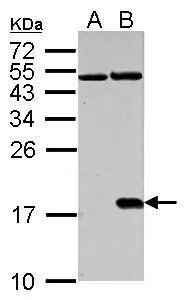
Histone H2A.X (phospho Ser139) antibody detects H2AFX protein in cisplatin-treated samples by western blot analysis. A. 30 μg NIH-3T3 whole cell lysate/extract (untreated) B. 30 μg NIH-3T3 whole cell lysate/extract (30μM cisplatin treatment for 24hr) 15% SDS-PAGE Histone H2A.X (phospho Ser139) antibody (GTX127340) dilution: 1:1000 The HRP-conjugated anti-rabbit IgG antibody (GTX213110-01) was used to detect the primary antibody.
 antibody detects H2AFX protein by western blot analysis. A. 30 μg HCT116 whole cell lysate/extract (untreated for 2hr) B. 30 μg HCT116 whole cell lysate/extract (UVB treatment 50mJ for 2hr) C. 30 μg HCT116 whole cell lysate/extract (untreated for 6hr) D. 30 μg HCT116 whole cell lysate/extract (UVB treatment 50mJ for 6hr) 15% SDS-PAGE Histone H2A.X (phospho Ser139) antibody (GTX127340) dilution: 1:1000 The HRP-conjugated anti-rabbit IgG antibody (GTX213110-01) was used to detect the primary antibody.")
 antibody detects Histone H2A.XS139ph (phospho Ser139) protein at nucleus by immunofluorescent analysis. Sample: HeLa cells were fixed in 4% paraformaldehyde at RT for 15 min. Green: Histone H2A.XS139ph (phospho Ser139) stained by Histone H2A.XS139ph (phospho Ser139) antibody (GTX127340) diluted at 1:1000. Red: phalloidin, a cytoskeleton marker, diluted at 1:100. Scale bar= 10 μm.")
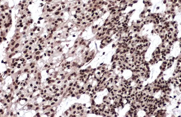
 antibody detects Histone H2A.XS139ph (phospho Ser139) protein at nucleus by immunohistochemical analysis. Sample: Paraffin-embedded human breast carcinoma. Histone H2A.XS139ph (phospho Ser139) stained by Histone H2A.XS139ph (phospho Ser139) antibody (GTX127340) diluted at 1:500. Antigen Retrieval: Citrate buffer, pH 6.0, 15 min")
 and treated (+) PC-12 whole cell extracts (30 μg) were separated by 15% SDS-PAGE, and the membrane was blotted with Histone H2A.XS139ph (phospho Ser139) antibody (GTX127340) diluted at 1:1000. The HRP-conjugated anti-rabbit IgG antibody (GTX213110-01) was used to detect the primary antibody.")
 and treated (+) HCT-116 whole cell extracts (30 μg) were separated by 15% SDS-PAGE, and the membrane was blotted with Histone H2A.XS139ph (phospho Ser139) antibody (GTX127340) diluted at 1:1000. The HRP-conjugated anti-rabbit IgG antibody (GTX213110-01) was used to detect the primary antibody.")


Histone H2A.X (phospho Ser139) antibody detects H2AFX protein in cisplatin-treated samples by western blot analysis. A. 30 μg NIH-3T3 whole cell lysate/extract (untreated) B. 30 μg NIH-3T3 whole cell lysate/extract (30μM cisplatin treatment for 24hr) 15% SDS-PAGE Histone H2A.X (phospho Ser139) antibody (GTX127340) dilution: 1:1000 The HRP-conjugated anti-rabbit IgG antibody (GTX213110-01) was used to detect the primary antibody.
Histone H2A.XS139ph (phospho Ser139) antibody
GTX127340
ApplicationsImmunoFluorescence, Western Blot, ImmunoCytoChemistry, ImmunoHistoChemistry, ImmunoHistoChemistry Frozen, ImmunoHistoChemistry Paraffin
Product group Antibodies
ReactivityHuman, Mouse, Rat, Zebra Fish
TargetH2AX
Overview
- SupplierGeneTex
- Product NameHistone H2A.XS139ph (phospho Ser139) antibody
- Delivery Days Customer9
- Application Supplier NoteWB: 1:500-1:3000. ICC/IF: 1:100-1:1000. IHC-P: 1:100-1:1000. *Optimal dilutions/concentrations should be determined by the researcher.Not tested in other applications.
- ApplicationsImmunoFluorescence, Western Blot, ImmunoCytoChemistry, ImmunoHistoChemistry, ImmunoHistoChemistry Frozen, ImmunoHistoChemistry Paraffin
- CertificationResearch Use Only
- ClonalityPolyclonal
- Concentration0.16 mg/ml
- ConjugateUnconjugated
- Gene ID3014
- Target nameH2AX
- Target descriptionH2A.X variant histone
- Target synonymsH2A.X, H2A/X, H2AFX, histone H2AX, H2A histone family member X, H2AX histone, histone H2A.x
- HostRabbit
- IsotypeIgG
- Protein IDP16104
- Protein NameHistone H2AX
- Scientific DescriptionHistones are basic nuclear proteins that are responsible for the nucleosome structure of the chromosomal fiber in eukaryotes. Two molecules of each of the four core histones (H2A, H2B, H3, and H4) form an octamer, around which approximately 146 bp of DNA is wrapped in repeating units, called nucleosomes. The linker histone, H1, interacts with linker DNA between nucleosomes and functions in the compaction of chromatin into higher order structures. This gene encodes a member of the histone H2A family, and generates two transcripts through the use of the conserved stem-loop termination motif, and the polyA addition motif. [provided by RefSeq]
- ReactivityHuman, Mouse, Rat, Zebra Fish
- Storage Instruction-20°C or -80°C,2°C to 8°C
- UNSPSC12352203
References
- Giang LH, Wu KS, Lee WC, et al. Targeting of RRM2 suppresses DNA damage response and activates apoptosis in atypical teratoid rhabdoid tumor. J Exp Clin Cancer Res. 2023,42(1):346. doi: 10.1186/s13046-023-02911-xRead this paper
- Jeong H, Wie M, Baek IJ, et al. TRIP13 Participates in Immediate-Early Sensing of DNA Strand Breaks and ATM Signaling Amplification through MRE11. Cells. 2022,11(24). doi: 10.3390/cells11244095Read this paper
- Zhou X, Zhou Q, Zhou D, et al. MicroRNA expression profiles in extracellular vesicles and intracellular of AURKA inhibitor-induced senescent neuroblastoma cells. Transl Cancer Res. 2022,11(8):2767-2782. doi: 10.21037/tcr-21-2438Read this paper
- Shih HT, Chen WY, Wang HY, et al. DNMT3b protects centromere integrity by restricting R-loop-mediated DNA damage. Cell Death Dis. 2022,13(6):546. doi: 10.1038/s41419-022-04989-1Read this paper
- Huang S, Wang X, Lin G, et al. Discovery of human TyrRS inhibitors by structure-based virtual screening, structural optimization, and bioassays. RSC Adv. 2019,9(16):9323-9330. doi: 10.1039/c9ra00458kRead this paper
- Yang JL, Yang MD, Chen JC, et al. Ouabain Induces DNA Damage in Human Osteosarcoma U-2 OS Cells and Alters the Expression of DNA Damage and DNA Repair-associated Proteins. In Vivo. 2021,35(5):2687-2696. doi: 10.21873/invivo.12552Read this paper
- Vanhunsel S, Bergmans S, Beckers A, et al. The killifish visual system as an in vivo model to study brain aging and rejuvenation. NPJ Aging Mech Dis. 2021,7(1):22. doi: 10.1038/s41514-021-00077-4Read this paper
- Cheng YC, Chen PY, Way TD, et al. Pre-Treatment of Pterostilbene Enhances H(2)O(2)-induced Cell Apoptosis Through Caspase-dependent Pathway in Human Keratinocyte Cells. In Vivo. 2021,35(2):833-843. doi: 10.21873/invivo.12324Read this paper
- Zhao T, Ye S, Tang Z, et al. Loss-of-function of p53 isoform Δ113p53 accelerates brain aging in zebrafish. Cell Death Dis. 2021,12(2):151. doi: 10.1038/s41419-021-03438-9Read this paper
- Chao T, Shih HT, Hsu SC, et al. Autophagy restricts mitochondrial DNA damage-induced release of ENDOG (endonuclease G) to regulate genome stability. Autophagy. 2021,17(11):3444-3460. doi: 10.1080/15548627.2021.1874209Read this paper
Datasheet
Related products
Product group Antibodies
ApplicationsDot Blot, Western Blot, ELISA
ReactivityHuman
TargetH2AX
- SizePrice
Product group Antibodies
Anti-Histone H2AX Antibody144-65468
ApplicationsImmunoFluorescence, Western Blot, ImmunoHistoChemistry
ReactivityHuman, Mouse, Rat
TargetH2AX
- SizePrice

Product group Antibodies
Anti-Histone H2A.X/H2AFX Antibody Picoband(r)A00241-1-CARRIER-FREE
ApplicationsFlow Cytometry, ImmunoPrecipitation, Western Blot, ELISA, ImmunoHistoChemistry
ReactivityHuman, Mouse, Rat
TargetH2AX
- SizePrice
Product group Antibodies
References
Histone H2A.X antibodyGTX108272
ApplicationsImmunoFluorescence, ImmunoPrecipitation, Western Blot, ImmunoCytoChemistry, ImmunoHistoChemistry, ImmunoHistoChemistry Paraffin
ReactivityHuman, Mouse, Rat
TargetH2AX
- SizePrice
![Histone H2A.X antibody [N1N2], N-term detects Histone H2A.X protein at nucleus by immunofluorescent analysis. Sample: A549 cells were fixed in 4% paraformaldehyde at RT for 15 min. Green: Histone H2A.X protein stained by Histone H2A.X antibody [N1N2], N-term (GTX108297) diluted at 1:500. Blue: Hoechst 33342 staining. Scale bar = 10 μm.](https://www.genetex.com/upload/website/prouct_img/normal/GTX108297/GTX108297_39750_20141205_IFA_w_23060120_599.webp)
Product group Antibodies
ApplicationsImmunoFluorescence, Western Blot, ImmunoCytoChemistry, ImmunoHistoChemistry, ImmunoHistoChemistry Paraffin
ReactivityHuman
TargetH2AX
- SizePrice
![WB analysis of A375, HEK293, HeLa and SK-MEL-2 whole cell lysates using GTX60914 Histone H2A.X antibody [RM214]. Dilution : 0.5μg/ml](https://www.genetex.com/upload/website/prouct_img/normal/GTX60914/GTX60914_20200909_WB_253_w_23061123_238.webp)
Product group Antibodies
Histone H2A.X antibody [RM214]GTX60914
ApplicationsImmunoFluorescence, Western Blot, ELISA, ImmunoCytoChemistry
ReactivityHuman
TargetH2AX
- SizePrice
![Untreated (–) and treated (+) HCT116 whole cell extracts (30 μg) were separated by 12% SDS-PAGE, and the membrane was blotted with Histone H2A.XS139ph (phospho Ser139) antibody [GT2311] (GTX628789) diluted at 1:1000. The HRP-conjugated anti-mouse IgG antibody (GTX213111-01) was used to detect the primary antibody, and the signal was developed with Trident ECL plus-Enhanced.](https://www.genetex.com/upload/website/prouct_img/normal/GTX628789/GTX628789_41190_20221216_WB_treatment_cisplatin_22122018_295.webp)
Product group Antibodies
References
ApplicationsImmunoFluorescence, ImmunoPrecipitation, Western Blot, ImmunoCytoChemistry, ImmunoHistoChemistry, ImmunoHistoChemistry Paraffin
ReactivityHuman, Mouse, Rat
TargetH2AX
- SizePrice
![Histone H2A.XS139ph (phospho Ser139) antibody [GT1021] detects Histone H2A.XS139ph (phospho Ser139) protein at Nucleus by immunofluorescent analysis. Sample: 10uM Cisplatin for 24 hrs HeLa cells were fixed in 4% paraformaldehyde/PBS for 15 min. Green: Histone H2A.XS139ph (phospho Ser139) protein stained by Histone H2A.XS139ph (phospho Ser139) antibody [GT1021] (GTX628996) diluted at 1:500. Blue: Hoechst 33342 staining.](https://www.genetex.com/upload/website/prouct_img/normal/GTX628996/GTX628996_41288_IFA_w_23061202_265.webp)
Product group Antibodies
References
ApplicationsImmunoFluorescence, Western Blot, ImmunoCytoChemistry
ReactivityHuman, Mouse
TargetH2AX
- SizePrice
![Histone H2A.XS139ph (phospho Ser139) antibody [HL1299] detects Histone H2A.XS139ph (phospho Ser139) protein at nucleus by immunohistochemical analysis. Sample: Paraffin-embedded mouse testis. Histone H2A.XS139ph (phospho Ser139) stained by Histone H2A.XS139ph (phospho Ser139) antibody [HL1299] (GTX636713) diluted at 1:100. Antigen Retrieval: Citrate buffer, pH 6.0, 15 min](https://www.genetex.com/upload/website/prouct_img/normal/GTX636713/GTX636713_44620_20220408_IHC-P_M_22071401_185.webp)
Product group Antibodies
ApplicationsImmunoFluorescence, Western Blot, ImmunoCytoChemistry, ImmunoHistoChemistry, ImmunoHistoChemistry Paraffin
ReactivityHuman, Mouse
TargetH2AX
- SizePrice
![ICC/IF analysis of cells using GTX80694 Histone H2A.XS139ph (phospho Ser139) antibody [3F2].](https://www.genetex.com/upload/website/prouct_img/normal/GTX80694/GTX80694_2034_ICC-IF_w_23061322_855.webp)
Product group Antibodies
References
ApplicationsFlow Cytometry, ImmunoFluorescence, Western Blot, ELISA, ImmunoCytoChemistry, ImmunoHistoChemistry, ImmunoHistoChemistry Paraffin
ReactivityBovine, Human, Mouse
TargetH2AX
- SizePrice