

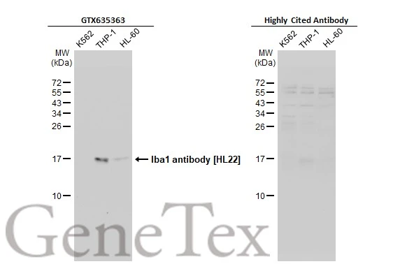
Various whole cell extracts (30 μg) were separated by 15% SDS-PAGE, and the membranes were blotted with Iba1 antibody [HL22] (GTX635363) diluted at 1:1000 and competitor's antibody diluted at 1:1000. The HRP-conjugated anti-rabbit IgG antibody (GTX213110-01) was used to detect the primary antibody, and the signal was developed with Trident ECL plus-Enhanced. *The competitor is not affiliated with GeneTex and does not endorse this product.
![Iba1 antibody [HL22] detects Iba1 protein by immunohistochemical analysis. Sample: Paraffin-embedded rat cerebellum. Iba1 stained by Iba1 antibody [HL22] (GTX635363) diluted at 1:1000. Antigen Retrieval: Citrate buffer, pH 6.0, 15 min](https://www.genetex.com/upload/website/prouct_img/normal/GTX635363/GTX635363_20200217_IHC-P_R_competitor_w_23061202_211.webp "Iba1 antibody [HL22] detects Iba1 protein by immunohistochemical analysis. Sample: Paraffin-embedded rat cerebellum. Iba1 stained by Iba1 antibody [HL22] (GTX635363) diluted at 1:1000. Antigen Retrieval: Citrate buffer, pH 6.0, 15 min")
![Iba1 antibody [HL22] detects Iba1 protein by immunohistochemical analysis. Sample: Frozen-sectioned mouse spinal cord. Green: Iba1 stained by Iba1 antibody [HL22] (GTX635363) diluted at 1:200. Blue: Fluoroshield with DAPI (GTX30920).](https://www.genetex.com/upload/website/prouct_img/normal/GTX635363/GTX635363_20200217_IHC-Fr_M_competitor_w_23061202_203.webp "Iba1 antibody [HL22] detects Iba1 protein by immunohistochemical analysis. Sample: Frozen-sectioned mouse spinal cord. Green: Iba1 stained by Iba1 antibody [HL22] (GTX635363) diluted at 1:200. Blue: Fluoroshield with DAPI (GTX30920).")
![Iba1 antibody [HL22] detects Iba1 protein by immunohistochemical analysis. Sample: Frozen-sectioned mouse brain. Green: Iba1 stained by Iba1 antibody [HL22] (GTX635363) diluted at 1:100. Red: Iba1 stained by Iba1 antibody [HL22-RT] (GTX635400) diluted at 1:100. Blue: Fluoroshield with DAPI (GTX30920).](https://www.genetex.com/upload/website/prouct_img/normal/GTX635363/GTX635363_43880_20200526_IHC-Fr_M_w_23061202_240.webp "Iba1 antibody [HL22] detects Iba1 protein by immunohistochemical analysis. Sample: Frozen-sectioned mouse brain. Green: Iba1 stained by Iba1 antibody [HL22] (GTX635363) diluted at 1:100. Red: Iba1 stained by Iba1 antibody [HL22-RT] (GTX635400) diluted at 1:100. Blue: Fluoroshield with DAPI (GTX30920).")
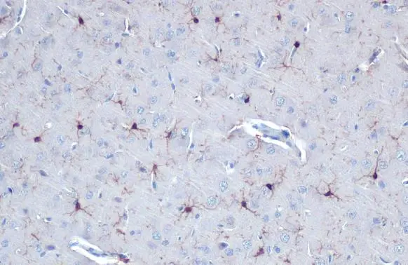
![Iba1 antibody [HL22] detects Iba1 protein by immunohistochemical analysis. Sample: Paraffin-embedded mouse hippocampus. Iba1 stained by Iba1 antibody [HL22] (GTX635363) diluted at 1:1000. Antigen Retrieval: Citrate buffer, pH 6.0, 15 min](https://www.genetex.com/upload/website/prouct_img/normal/GTX635363/GTX635363_20200217_IHC-P_M_competitor_w_23061202_824.webp "Iba1 antibody [HL22] detects Iba1 protein by immunohistochemical analysis. Sample: Paraffin-embedded mouse hippocampus. Iba1 stained by Iba1 antibody [HL22] (GTX635363) diluted at 1:1000. Antigen Retrieval: Citrate buffer, pH 6.0, 15 min")
![Iba1 antibody [HL22] detects Iba1 protein at cytoplasm by immunofluorescent analysis. Sample: THP-1 cells were fixed in 4% paraformaldehyde at RT for 15 min. Green: Iba1 stained by Iba1 antibody [HL22] (GTX635363) diluted at 1:500. Blue: Hoechst 33342 staining.](https://www.genetex.com/upload/website/prouct_img/normal/GTX635363/GTX635363_44599_20220415_ICC_IF_w_23061202_877.webp "Iba1 antibody [HL22] detects Iba1 protein at cytoplasm by immunofluorescent analysis. Sample: THP-1 cells were fixed in 4% paraformaldehyde at RT for 15 min. Green: Iba1 stained by Iba1 antibody [HL22] (GTX635363) diluted at 1:500. Blue: Hoechst 33342 staining.")
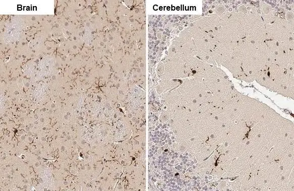
![Iba1 antibody [HL22] detects Iba1 protein at cell membrane and cytoplasm by immunohistochemical analysis. Sample: Paraffin-embedded rat brain. Iba1 stained by Iba1 antibody [HL22] (GTX635363) diluted at 1:100. Antigen Retrieval: Citrate buffer, pH 6.0, 15 min](https://www.genetex.com/upload/website/prouct_img/normal/GTX635363/GTX635363_44403_20210820_IHC-P_R_w_23061202_407.webp "Iba1 antibody [HL22] detects Iba1 protein at cell membrane and cytoplasm by immunohistochemical analysis. Sample: Paraffin-embedded rat brain. Iba1 stained by Iba1 antibody [HL22] (GTX635363) diluted at 1:100. Antigen Retrieval: Citrate buffer, pH 6.0, 15 min")
![Iba1 antibody [HL22] detects Iba1 protein by immunohistochemical analysis. Sample: Paraffin-embedded rat brain. Iba1 stained by Iba1 antibody [HL22] (GTX635363) conjugated with DyLight488 diluted at 1:200. Blue: Fluoroshield with DAPI (GTX30920). Antigen Retrieval: Citrate buffer, pH 6.0, 15 min.](https://www.genetex.com/upload/website/prouct_img/normal/GTX635363/GTX635363_20220509_IHC-P_R_w_23061202_504.webp "Iba1 antibody [HL22] detects Iba1 protein by immunohistochemical analysis. Sample: Paraffin-embedded rat brain. Iba1 stained by Iba1 antibody [HL22] (GTX635363) conjugated with DyLight488 diluted at 1:200. Blue: Fluoroshield with DAPI (GTX30920). Antigen Retrieval: Citrate buffer, pH 6.0, 15 min.")
![Various whole cell extracts (30 μg) were separated by 15% SDS-PAGE, and the membrane was blotted with Iba1 antibody [HL22] (GTX635363) diluted at 1:1000. The HRP-conjugated anti-rabbit IgG antibody (GTX213110-01) was used to detect the primary antibody. Corresponding RNA expression data for the same cell lines are based on Human Protein Atlas program.](https://www.genetex.com/upload/website/prouct_img/normal/GTX635363/GTX635363_45251_20231208_WB_TPM_watermark_24071520_593.webp "Various whole cell extracts (30 μg) were separated by 15% SDS-PAGE, and the membrane was blotted with Iba1 antibody [HL22] (GTX635363) diluted at 1:1000. The HRP-conjugated anti-rabbit IgG antibody (GTX213110-01) was used to detect the primary antibody. Corresponding RNA expression data for the same cell lines are based on Human Protein Atlas program.")
Various whole cell extracts (30 μg) were separated by 15% SDS-PAGE, and the membranes were blotted with Iba1 antibody [HL22] (GTX635363) diluted at 1:1000 and competitor's antibody diluted at 1:1000. The HRP-conjugated anti-rabbit IgG antibody (GTX213110-01) was used to detect the primary antibody, and the signal was developed with Trident ECL plus-Enhanced. *The competitor is not affiliated with GeneTex and does not endorse this product.
Iba1 antibody [HL22]
GTX635363
ApplicationsImmunoFluorescence, Western Blot, ImmunoCytoChemistry, ImmunoHistoChemistry, ImmunoHistoChemistry Frozen, ImmunoHistoChemistry Paraffin
Product group Antibodies
ReactivityHuman, Mouse, Porcine, Rat
TargetAIF1
Overview
- SupplierGeneTex
- Product NameIba1 antibody [HL22]
- Delivery Days Customer9
- Application Supplier NoteWB: 1:500-1:3000. IHC-P: 1:100-1:1000. IHC-Fr: 1:100-1:1000. *Optimal dilutions/concentrations should be determined by the researcher.Not tested in other applications.
- ApplicationsImmunoFluorescence, Western Blot, ImmunoCytoChemistry, ImmunoHistoChemistry, ImmunoHistoChemistry Frozen, ImmunoHistoChemistry Paraffin
- CertificationResearch Use Only
- ClonalityMonoclonal
- Clone IDHL22
- Concentration1 mg/ml
- ConjugateUnconjugated
- Gene ID199
- Target nameAIF1
- Target descriptionallograft inflammatory factor 1
- Target synonymsAIF-1, IBA1, IRT-1, IRT1, allograft inflammatory factor 1, interferon gamma responsive transcript, ionized calcium-binding adapter molecule 1, protein G1
- HostRabbit
- IsotypeIgG
- Protein IDP55008
- Protein NameAllograft inflammatory factor 1
- Scientific DescriptionThis gene encodes a protein that binds actin and calcium. This gene is induced by cytokines and interferon and may promote macrophage activation and growth of vascular smooth muscle cells and T-lymphocytes. Polymorphisms in this gene may be associated with systemic sclerosis. Alternative splicing results in multiple transcript variants, but the full-length and coding nature of some of these variants is not certain. [provided by RefSeq, Jan 2016]
- ReactivityHuman, Mouse, Porcine, Rat
- Storage Instruction-20°C or -80°C,2°C to 8°C
- UNSPSC12352203
References
- Iacono D, Murphy EK, Stimpson CD, et al. Low-dose brain radiation: lowering hyperphosphorylated-tau without increasing DNA damage or oncogenic activation. Sci Rep. 2023,13(1):21142. doi: 10.1038/s41598-023-48146-wRead this paper
- Brown TC, Crouse EC, Attaway CA, et al. Microglia are dispensable for experience-dependent refinement of visual circuitry. bioRxiv. 2023,:pii: 2023.10.17.562708. doi: 10.1101/2023.10.17.562708.Read this paper
- Wang HK, Su YT, Ho YC, et al. HDAC1 is Involved in Neuroinflammation and Blood-Brain Barrier Damage in Stroke Pathogenesis. J Inflamm Res. 2023,16:4103-4116. doi: 10.2147/JIR.S416239Read this paper
- Wong CE, Hu CY, Lee PH, et al. Sciatic nerve stimulation alleviates acute neuropathic pain via modulation of neuroinflammation and descending pain inhibition in a rodent model. J Neuroinflammation. 2022,19(1):153. doi: 10.1186/s12974-022-02513-yRead this paper
- Fan Z, Zhang W, Cao Q, et al. JAK2/STAT3 pathway regulates microglia polarization involved in hippocampal inflammatory damage due to acute paraquat exposure. Ecotoxicol Environ Saf. 2022,234:113372. doi: 10.1016/j.ecoenv.2022.113372Read this paper
- Zhang Y, Hou B, Liang P, et al. TRPV1 channel mediates NLRP3 inflammasome-dependent neuroinflammation in microglia. Cell Death Dis. 2021,12(12):1159. doi: 10.1038/s41419-021-04450-9Read this paper
- Yan H, Zhao H, Kang Y, et al. Parecoxib alleviates the motor behavioral decline of aged rats by ameliorating mitochondrial dysfunction in the substantia nigra via COX-2/PGE2 pathway inhibition. Neuropharmacology. 2021,194:108627. doi: 10.1016/j.neuropharm.2021.108627Read this paper
- Xiong LL, Tan YX, Du RL, et al. Effect of Sutellarin on Neurogenesis in Neonatal Hypoxia-Ischemia Rat Model: Potential Mechanisms of Action. Am J Chin Med. 2021,49(3):677-703. doi: 10.1142/S0192415X21500312Read this paper
- Teng SW, Sung HY, Wen YC, et al. Potential surrogate quantitative immunomodulatory potency assay for monitoring human umbilical cord-derived mesenchymal stem cells production. Cell Biol Int. 2021,45(5):1072-1081. doi: 10.1002/cbin.11553Read this paper
Datasheet
Related products
Product group Antibodies

Anti-AIF1 [GT1-mAb1]AB03986-1.1
ApplicationsFlow Cytometry, ImmunoFluorescence, Western Blot, ELISA
ReactivityHuman
TargetAIF1
- SizePrice
Product group Antibodies
Anti-AIF1 Antibody144-60080
ApplicationsWestern Blot
ReactivityHuman, Mouse, Rat
TargetAIF1
- SizePrice

Product group Antibodies
Anti-Iba1/AIF1 Antibody Picoband(r)A01394-CARRIER-FREE
ApplicationsWestern Blot, ImmunoHistoChemistry
ReactivityHuman
TargetAIF1
- SizePrice
![IHC-P analysis of human placenta tissue using GTX04465 Iba1 antibody [MSVA-955M] HistoMAX?. Early Iba1 expression is particularly strong in macrophages of the placenta.](https://www.genetex.com/upload/website/prouct_img/normal/GTX04465/GTX04465_20230728_IHC-P_1_23072722_931.webp)
Product group Antibodies
ApplicationsImmunoHistoChemistry, ImmunoHistoChemistry Paraffin
ReactivityHuman
TargetAIF1
- SizePrice
Product group Antibodies
References
Iba1 antibodyGTX100042
ApplicationsFlow Cytometry, ImmunoFluorescence, Western Blot, ImmunoCytoChemistry, ImmunoHistoChemistry, ImmunoHistoChemistry Frozen, ImmunoHistoChemistry Paraffin
ReactivityHuman, Mouse, Rat
TargetAIF1
- SizePrice
Product group Antibodies
References
Iba1 antibodyGTX101495
ApplicationsFlow Cytometry, ImmunoFluorescence, ImmunoPrecipitation, Western Blot, ImmunoCytoChemistry, ImmunoHistoChemistry, ImmunoHistoChemistry Frozen, ImmunoHistoChemistry Paraffin
ReactivityHuman, Mouse, Rat
TargetAIF1
- SizePrice
![Whole cell extract (30 μg) was separated by 15% SDS-PAGE, and the membrane was blotted with Iba1 antibody [GT10312] (GTX632426) diluted at 1:1000. The HRP-conjugated anti-mouse IgG antibody (GTX213111-01) was used to detect the primary antibody, and the signal was developed with Trident ECL plus-Enhanced.](https://www.genetex.com/upload/website/prouct_img/normal/GTX632426/GTX632426_44825_20230324_WB_Z_brain_23032819_167.webp)
Product group Antibodies
References
Iba1 antibody [GT10312]GTX632426
ApplicationsFlow Cytometry, ImmunoFluorescence, Western Blot, ImmunoCytoChemistry, ImmunoHistoChemistry, ImmunoHistoChemistry Paraffin
ReactivityHuman, Mouse, Rat, Zebra Fish
TargetAIF1
- SizePrice
![Iba1 antibody [HL22-MS] detects Iba1 protein at cell membrane and cytoplasm by immunohistochemical analysis. Sample: Frozen-sectioned mouse brain. Green: Iba1 stained by Iba1 antibody [HL22-MS] (GTX635399) diluted at 1:25. Blue: Fluoroshield with DAPI (GTX30920).](https://www.genetex.com/upload/website/prouct_img/normal/GTX635399/GTX635399_43917_20211022_IHC-Fr_M_w_23061202_468.webp)
Product group Antibodies
References
Iba1 antibody [HL22-MS]GTX635399
ApplicationsWestern Blot, ImmunoHistoChemistry, ImmunoHistoChemistry Frozen, ImmunoHistoChemistry Paraffin
ReactivityHuman, Mouse, Rat
TargetAIF1
- SizePrice
![Iba1 antibody [HL22-RT] detects Iba1 protein at cytoplasm by immunofluorescent analysis. Sample: THP-1 cells were fixed in 4% paraformaldehyde at RT for 15 min. Green: Iba1 stained by Iba1 antibody [HL22-RT] (GTX635400) diluted at 1:500. Blue: Fluoroshield with DAPI (GTX30920).](https://www.genetex.com/upload/website/prouct_img/normal/GTX635400/GTX635400_44858_20230331_ICC_IF_23041023_641.webp)
Product group Antibodies
Iba1 antibody [HL22-RT]GTX635400
ApplicationsImmunoFluorescence, Western Blot, ImmunoCytoChemistry, ImmunoHistoChemistry, ImmunoHistoChemistry Frozen, ImmunoHistoChemistry Paraffin
ReactivityHuman, Mouse, Rat
TargetAIF1
- SizePrice