

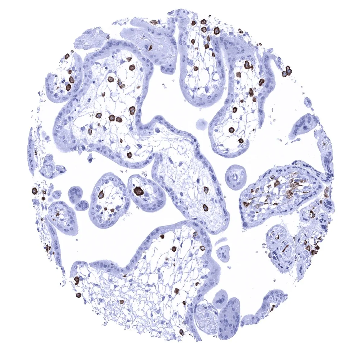
IHC-P analysis of human placenta tissue using GTX04465 Iba1 antibody [MSVA-955M] HistoMAX?. Early Iba1 expression is particularly strong in macrophages of the placenta.
![IHC-P analysis of human mucosa from duodenum tissue using GTX04465 Iba1 antibody [MSVA-955M] HistoMAX?. A strong Iba1 positivity is seen in mucosal macrophages of the duodenum.](https://www.genetex.com/upload/website/prouct_img/normal/GTX04465/GTX04465_20230728_IHC-P_131_23072722_660.webp "IHC-P analysis of human mucosa from duodenum tissue using GTX04465 Iba1 antibody [MSVA-955M] HistoMAX?. A strong Iba1 positivity is seen in mucosal macrophages of the duodenum.")
![IHC-P analysis of human epithelioid malignant mesothelioma tissue using GTX04465 Iba1 antibody [MSVA-955M] HistoMAX?. Malignant mesothelioma epitheloid subtype containing numerous Iba1 positive inflammatory cells.](https://www.genetex.com/upload/website/prouct_img/normal/GTX04465/GTX04465_20230728_IHC-P_261_23072723_120.webp "IHC-P analysis of human epithelioid malignant mesothelioma tissue using GTX04465 Iba1 antibody [MSVA-955M] HistoMAX?. Malignant mesothelioma epitheloid subtype containing numerous Iba1 positive inflammatory cells.")
IHC-P analysis of human placenta tissue using GTX04465 Iba1 antibody [MSVA-955M] HistoMAX?. Early Iba1 expression is particularly strong in macrophages of the placenta.
Iba1 antibody [MSVA-955M] HistoMAX(tm)
GTX04465
ApplicationsImmunoHistoChemistry, ImmunoHistoChemistry Paraffin
Product group Antibodies
ReactivityHuman
TargetAIF1
Overview
- SupplierGeneTex
- Product NameIba1 antibody [MSVA-955M] HistoMAX(tm)
- Delivery Days Customer9
- Application Supplier NoteIHC-P: 1:100-1:200. *Optimal dilutions/concentrations should be determined by the researcher.Not tested in other applications.
- ApplicationsImmunoHistoChemistry, ImmunoHistoChemistry Paraffin
- CertificationResearch Use Only
- ClonalityMonoclonal
- Clone IDMSVA-955M
- Concentration0.2 mg/ml
- ConjugateUnconjugated
- Gene ID199
- Target nameAIF1
- Target descriptionallograft inflammatory factor 1
- Target synonymsAIF-1, IBA1, IRT-1, IRT1, allograft inflammatory factor 1, interferon gamma responsive transcript, ionized calcium-binding adapter molecule 1, protein G1
- HostMouse
- IsotypeIgG2c
- Protein IDP55008
- Protein NameAllograft inflammatory factor 1
- Scientific DescriptionThis gene encodes a protein that binds actin and calcium. This gene is induced by cytokines and interferon and may promote macrophage activation and growth of vascular smooth muscle cells and T-lymphocytes. Polymorphisms in this gene may be associated with systemic sclerosis. Alternative splicing results in multiple transcript variants, but the full-length and coding nature of some of these variants is not certain. [provided by RefSeq, Jan 2016]
- ReactivityHuman
- Storage Instruction-20°C or -80°C,2°C to 8°C
- UNSPSC12352203
Datasheet
Related products
Product group Antibodies

Anti-AIF1 [GT1-mAb1]AB03986-1.1
ApplicationsFlow Cytometry, ImmunoFluorescence, Western Blot, ELISA
ReactivityHuman
TargetAIF1
- SizePrice
Product group Antibodies
Anti-AIF1 Antibody144-60080
ApplicationsWestern Blot
ReactivityHuman, Mouse, Rat
TargetAIF1
- SizePrice

Product group Antibodies
Anti-Iba1/AIF1 Antibody Picoband(r)A01394-CARRIER-FREE
ApplicationsWestern Blot, ImmunoHistoChemistry
ReactivityHuman
TargetAIF1
- SizePrice
Product group Antibodies
References
Iba1 antibodyGTX100042
ApplicationsFlow Cytometry, ImmunoFluorescence, Western Blot, ImmunoCytoChemistry, ImmunoHistoChemistry, ImmunoHistoChemistry Frozen, ImmunoHistoChemistry Paraffin
ReactivityHuman, Mouse, Rat
TargetAIF1
- SizePrice
Product group Antibodies
References
Iba1 antibodyGTX101495
ApplicationsFlow Cytometry, ImmunoFluorescence, ImmunoPrecipitation, Western Blot, ImmunoCytoChemistry, ImmunoHistoChemistry, ImmunoHistoChemistry Frozen, ImmunoHistoChemistry Paraffin
ReactivityHuman, Mouse, Rat
TargetAIF1
- SizePrice
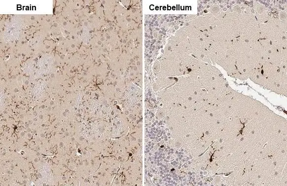
![Whole cell extract (30 μg) was separated by 15% SDS-PAGE, and the membrane was blotted with Iba1 antibody [GT10312] (GTX632426) diluted at 1:1000. The HRP-conjugated anti-mouse IgG antibody (GTX213111-01) was used to detect the primary antibody, and the signal was developed with Trident ECL plus-Enhanced.](https://www.genetex.com/upload/website/prouct_img/normal/GTX632426/GTX632426_44825_20230324_WB_Z_brain_23032819_167.webp)
Product group Antibodies
References
Iba1 antibody [GT10312]GTX632426
ApplicationsFlow Cytometry, ImmunoFluorescence, Western Blot, ImmunoCytoChemistry, ImmunoHistoChemistry, ImmunoHistoChemistry Paraffin
ReactivityHuman, Mouse, Rat, Zebra Fish
TargetAIF1
- SizePrice
![Various whole cell extracts (30 μg) were separated by 15% SDS-PAGE, and the membranes were blotted with Iba1 antibody [HL22] (GTX635363) diluted at 1:1000 and competitor's antibody diluted at 1:1000. The HRP-conjugated anti-rabbit IgG antibody (GTX213110-01) was used to detect the primary antibody, and the signal was developed with Trident ECL plus-Enhanced. *The competitor is not affiliated with GeneTex and does not endorse this product.](https://www.genetex.com/upload/website/prouct_img/normal/GTX635363/GTX635363_testlot22-5_20200217_WB_recAb_competitor_watermark_w_23061202_940.webp)
Product group Antibodies
References
Iba1 antibody [HL22]GTX635363
ApplicationsImmunoFluorescence, Western Blot, ImmunoCytoChemistry, ImmunoHistoChemistry, ImmunoHistoChemistry Frozen, ImmunoHistoChemistry Paraffin
ReactivityHuman, Mouse, Porcine, Rat
TargetAIF1
- SizePrice
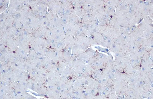
![Iba1 antibody [HL22-MS] detects Iba1 protein at cell membrane and cytoplasm by immunohistochemical analysis. Sample: Frozen-sectioned mouse brain. Green: Iba1 stained by Iba1 antibody [HL22-MS] (GTX635399) diluted at 1:25. Blue: Fluoroshield with DAPI (GTX30920).](https://www.genetex.com/upload/website/prouct_img/normal/GTX635399/GTX635399_43917_20211022_IHC-Fr_M_w_23061202_468.webp)
Product group Antibodies
References
Iba1 antibody [HL22-MS]GTX635399
ApplicationsWestern Blot, ImmunoHistoChemistry, ImmunoHistoChemistry Frozen, ImmunoHistoChemistry Paraffin
ReactivityHuman, Mouse, Rat
TargetAIF1
- SizePrice
![Iba1 antibody [HL22-RT] detects Iba1 protein at cytoplasm by immunofluorescent analysis. Sample: THP-1 cells were fixed in 4% paraformaldehyde at RT for 15 min. Green: Iba1 stained by Iba1 antibody [HL22-RT] (GTX635400) diluted at 1:500. Blue: Fluoroshield with DAPI (GTX30920).](https://www.genetex.com/upload/website/prouct_img/normal/GTX635400/GTX635400_44858_20230331_ICC_IF_23041023_641.webp)
Product group Antibodies
Iba1 antibody [HL22-RT]GTX635400
ApplicationsImmunoFluorescence, Western Blot, ImmunoCytoChemistry, ImmunoHistoChemistry, ImmunoHistoChemistry Frozen, ImmunoHistoChemistry Paraffin
ReactivityHuman, Mouse, Rat
TargetAIF1
- SizePrice