

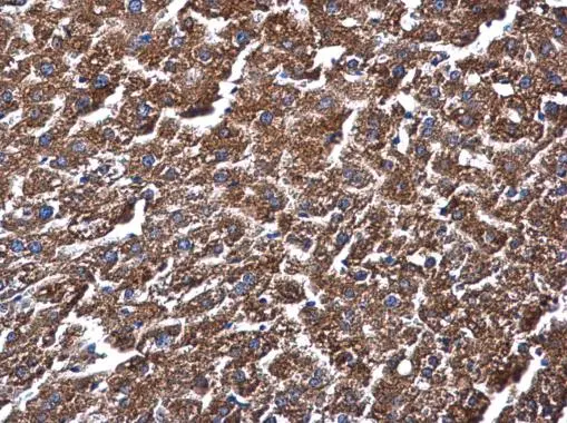
ME1 antibody [GT15611] detects ME1 protein at cytoplasm in mouse liver by immunohistochemical analysis. Sample: Paraffin-embedded mouse liver. ME1 antibody [GT15611] (GTX632190) diluted at 1:500.
Antigen Retrieval: Citrate buffer, pH 6.0, 15 min
![Double-labeled immunofluorescence photomicrographs of frozen sections of mouse brain. Green: Vimentin antibody (GTX100619) diluted at 1:200. The signal was developed using goat anti-rabbit IgG antibody (Dylight488) (GTX213110-04). Red: ME1 antibody [GT15611] (GTX632190) diluted at 1:200. The signal was developed using goat anti-mouse IgG antibody (Dylight594) (GTX213111-05). Blue: Nuclear staining with Hoechst 33342.
Antigen Retrieval: Citrate buffer, pH 6.0, 15 min](https://www.genetex.com/upload/website/prouct_img/normal/GTX632190/GTX632190_42037_20150814_IHC-Fr_M_w_23061202_630.webp "Double-labeled immunofluorescence photomicrographs of frozen sections of mouse brain. Green: Vimentin antibody (GTX100619) diluted at 1:200. The signal was developed using goat anti-rabbit IgG antibody (Dylight488) (GTX213110-04). Red: ME1 antibody [GT15611] (GTX632190) diluted at 1:200. The signal was developed using goat anti-mouse IgG antibody (Dylight594) (GTX213111-05). Blue: Nuclear staining with Hoechst 33342.
Antigen Retrieval: Citrate buffer, pH 6.0, 15 min")
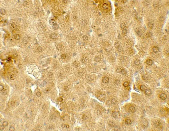
![ME1 antibody [GT15611] detects ME1 protein at cytoplasm in mouse cervix by immunohistochemical analysis. Sample: Paraffin-embedded mouse cervix. ME1 antibody [GT15611] (GTX632190) diluted at 1:500.
Antigen Retrieval: Citrate buffer, pH 6.0, 15 min](https://www.genetex.com/upload/website/prouct_img/normal/GTX632190/GTX632190_42037_20150625_IHC-P_M_w_23061202_154.webp "ME1 antibody [GT15611] detects ME1 protein at cytoplasm in mouse cervix by immunohistochemical analysis. Sample: Paraffin-embedded mouse cervix. ME1 antibody [GT15611] (GTX632190) diluted at 1:500.
Antigen Retrieval: Citrate buffer, pH 6.0, 15 min")
 were separated by 7.5% SDS-PAGE, and the membrane was blotted with ME1 antibody (GTX632190) diluted at a dilution of 1:1000.")
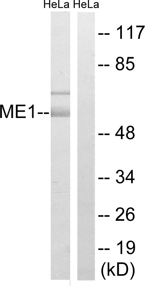
![Non-transfected (–) and transfected (+) HeLa whole cell extracts (30 μg) were separated by 7.5% SDS-PAGE, and the membrane was blotted with ME1 antibody [GT15611] (GTX632190) diluted at 1:1000.](https://www.genetex.com/upload/website/prouct_img/normal/GTX632190/GTX632190_42037_20160721_WB_shRNA_watermark_w_23061202_686.webp "Non-transfected (–) and transfected (+) HeLa whole cell extracts (30 μg) were separated by 7.5% SDS-PAGE, and the membrane was blotted with ME1 antibody [GT15611] (GTX632190) diluted at 1:1000.")
ME1 antibody [GT15611] detects ME1 protein at cytoplasm in mouse liver by immunohistochemical analysis. Sample: Paraffin-embedded mouse liver. ME1 antibody [GT15611] (GTX632190) diluted at 1:500.
Antigen Retrieval: Citrate buffer, pH 6.0, 15 min
ME1 antibody [GT15611]
GTX632190
ApplicationsWestern Blot, ImmunoHistoChemistry, ImmunoHistoChemistry Frozen, ImmunoHistoChemistry Paraffin
Product group Antibodies
ReactivityHuman, Mouse
TargetME1
Overview
- SupplierGeneTex
- Product NameME1 antibody [GT15611]
- Delivery Days Customer9
- Application Supplier NoteWB: 1:500-1:3000. IHC-P: 1:100-1:1000. IHC-Fr: 1:100-1:1000. *Optimal dilutions/concentrations should be determined by the researcher.Not tested in other applications.
- ApplicationsWestern Blot, ImmunoHistoChemistry, ImmunoHistoChemistry Frozen, ImmunoHistoChemistry Paraffin
- CertificationResearch Use Only
- ClonalityMonoclonal
- Clone IDGT15611
- Concentration1 mg/ml
- ConjugateUnconjugated
- Gene ID4199
- Target nameME1
- Target descriptionmalic enzyme 1
- Target synonymsHUMNDME, MES, NADP-dependent malic enzyme, Malic enzyme, cytoplasmic, NADP-ME, malate dehydrogenase (oxaloacetate-decarboxylating) (NADP(+)), malic enzyme 1, NADP(+)-dependent, cytosolic, malic enzyme 1, soluble, pyruvic-malic carboxylase
- HostMouse
- IsotypeIgG2a
- Protein IDP48163
- Protein NameNADP-dependent malic enzyme
- Scientific DescriptionThis gene encodes a cytosolic, NADP-dependent enzyme that generates NADPH for fatty acid biosynthesis. The activity of this enzyme, the reversible oxidative decarboxylation of malate, links the glycolytic and citric acid cycles. The regulation of expression for this gene is complex. Increased expression can result from elevated levels of thyroid hormones or by higher proportions of carbohydrates in the diet. [provided by RefSeq]
- ReactivityHuman, Mouse
- Storage Instruction-20°C or -80°C,2°C to 8°C
- UNSPSC41116161
Datasheet
Related products

Product group Antibodies
Anti-ME1 Antibody Picoband(r)A03449-3-CARRIER-FREE
ApplicationsFlow Cytometry, ImmunoFluorescence, Western Blot, ELISA, ImmunoCytoChemistry, ImmunoHistoChemistry
ReactivityHuman, Mouse, Rat
TargetME1
- SizePrice
Product group Antibodies
Anti-ME1 AntibodyA98477
ApplicationsWestern Blot, ELISA
ReactivityHuman, Mouse, Rat
- SizePrice

Product group Antibodies
Anti-ME1 AntibodyHPA006493
ApplicationsWestern Blot, ImmunoHistoChemistry
ReactivityHuman
TargetME1
- SizePrice
Product group Antibodies
ME1 AntibodyCSB-PA010149
ApplicationsWestern Blot, ELISA
ReactivityHuman, Mouse, Rat
TargetME1
- SizePrice
Product group Antibodies

ME1 / Malate Dehydrogenase AntibodyLS-C667898
ApplicationsWestern Blot
ReactivityHuman
TargetME1
- SizePrice
Product group Antibodies
ME1 antibodyGTX104122
ApplicationsImmunoFluorescence, ImmunoPrecipitation, Western Blot, ImmunoCytoChemistry, ImmunoHistoChemistry, ImmunoHistoChemistry Paraffin
ReactivityHuman, Mouse, Rat
TargetME1
- SizePrice
![Non-transfected (–) and transfected (+) HeLa whole cell extracts (30 μg) were separated by 7.5% SDS-PAGE, and the membrane was blotted with ME1 antibody [GT736] (GTX632188) diluted at 1:500.](https://www.genetex.com/upload/website/prouct_img/normal/GTX632188/GTX632188_42037_20160721_WB_shRNA_watermark_w_23061202_936.webp)
Product group Antibodies
ME1 antibody [GT736]GTX632188
ApplicationsWestern Blot
ReactivityHuman
TargetME1
- SizePrice
![ME1 antibody [GT979] detects ME1 protein at cytoplasm by immunofluorescent analysis. Sample: HeLa cells were fixed in 4% paraformaldehyde at RT for 15 min. Green: ME1 protein stained by ME1 antibody [GT979] (GTX632189) diluted at 1:400. Blue: Hoechst 33342 staining.](https://www.genetex.com/upload/website/prouct_img/normal/GTX632189/GTX632189_42037_20150831_IFA_w_23061202_318.webp)
Product group Antibodies
ME1 antibody [GT979]GTX632189
ApplicationsImmunoFluorescence, Western Blot, ImmunoCytoChemistry, ImmunoHistoChemistry, ImmunoHistoChemistry Paraffin
ReactivityHuman, Mouse
TargetME1
- SizePrice
Product group Antibodies
ME1 antibodyGTX31914
ApplicationsWestern Blot, ELISA, ImmunoHistoChemistry, ImmunoHistoChemistry Paraffin
ReactivityHuman, Mouse, Rat
TargetME1
- SizePrice