

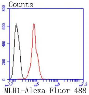
FACS analysis of HeLa cells using GTX01140 MLH1 antibody [SP08-04]. Red : primary antibody Black : unlabelled control Dilution : 1:50
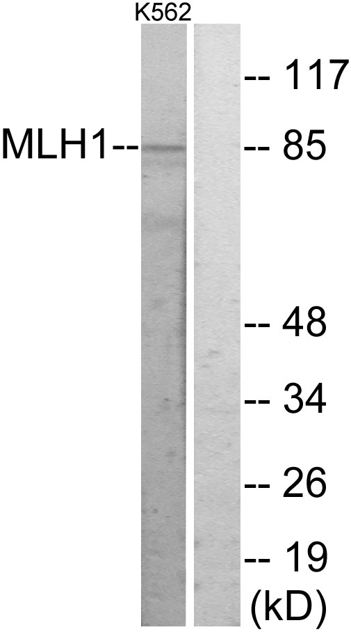
![Various whole cell extracts (30 μg) were separated by 7.5% SDS-PAGE, and the membrane was blotted with MLH1 antibody [SP08-04] (GTX01140) diluted at 1:500. The HRP-conjugated anti-rabbit IgG antibody (GTX213110-01) was used to detect the primary antibody.](https://www.genetex.com/upload/website/prouct_img/normal/GTX01140/GTX01140_HM0812_20200228_WB_w_23053121_521.webp "Various whole cell extracts (30 μg) were separated by 7.5% SDS-PAGE, and the membrane was blotted with MLH1 antibody [SP08-04] (GTX01140) diluted at 1:500. The HRP-conjugated anti-rabbit IgG antibody (GTX213110-01) was used to detect the primary antibody.")
FACS analysis of HeLa cells using GTX01140 MLH1 antibody [SP08-04]. Red : primary antibody Black : unlabelled control Dilution : 1:50
MLH1 antibody [SP08-04]
GTX01140
ApplicationsFlow Cytometry, Western Blot
Product group Antibodies
ReactivityHuman
TargetMLH1
Overview
- SupplierGeneTex
- Product NameMLH1 antibody [SP08-04]
- Delivery Days Customer9
- Application Supplier NoteWB: 1:1000. FCM: 1:50-1:100. *Optimal dilutions/concentrations should be determined by the researcher.Not tested in other applications.
- ApplicationsFlow Cytometry, Western Blot
- CertificationResearch Use Only
- ClonalityMonoclonal
- Clone IDSP08-04
- Concentration1 mg/ml
- ConjugateUnconjugated
- Gene ID4292
- Target nameMLH1
- Target descriptionmutL homolog 1
- Target synonymsCOCA2, FCC2, HNPCC, HNPCC2, LYNCH2, MLH-1, MMRCS1, hMLH1, DNA mismatch repair protein Mlh1, mutL homolog 1, colon cancer, nonpolyposis type 2
- HostRabbit
- IsotypeIgG
- Protein IDP40692
- Protein NameDNA mismatch repair protein Mlh1
- Scientific DescriptionThe protein encoded by this gene can heterodimerize with mismatch repair endonuclease PMS2 to form MutL alpha, part of the DNA mismatch repair system. When MutL alpha is bound by MutS beta and some accessory proteins, the PMS2 subunit of MutL alpha introduces a single-strand break near DNA mismatches, providing an entry point for exonuclease degradation. The encoded protein is also involved in DNA damage signaling and can heterodimerize with DNA mismatch repair protein MLH3 to form MutL gamma, which is involved in meiosis. This gene was identified as a locus frequently mutated in hereditary nonpolyposis colon cancer (HNPCC). [provided by RefSeq, Aug 2017]
- ReactivityHuman
- Storage Instruction-20°C or -80°C,2°C to 8°C
- UNSPSC41116161
Datasheet
Related products
Product group Antibodies
Anti-MLH1 AntibodyA98041
ApplicationsWestern Blot, ELISA
ReactivityHuman, Mouse, Rat
- SizePrice
Product group Antibodies
Anti-MLH1 Antibody118-10018
ApplicationsELISA, ImmunoHistoChemistry
ReactivityHuman
- SizePrice

Product group Antibodies
Anti-MLH1 Antibody Picoband(r)A00126-1-CARRIER-FREE
ApplicationsWestern Blot, ELISA, ImmunoHistoChemistry
ReactivityHuman
TargetMLH1
- SizePrice
Product group Antibodies
MLH1 Recombinant AntibodyBSM-60697R
ApplicationsWestern Blot
ReactivityHuman, Mouse
TargetMLH1
- SizePrice
Product group Antibodies
MLH1 AntibodyCSB-PA003247
ApplicationsImmunoFluorescence, Western Blot, ELISA
ReactivityHuman, Mouse, Rat
TargetMLH1
- SizePrice
Product group Antibodies
MLH1 Polyclonal AntibodyCAC14558
ApplicationsImmunoFluorescence, Western Blot, ELISA, ImmunoHistoChemistry
TargetMLH1
- SizePrice
Product group Antibodies

MLH1 AntibodyLS-C400894
ApplicationsELISA, ImmunoHistoChemistry
ReactivityHuman, Mouse, Rat
TargetMLH1
- SizePrice
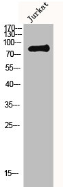
![WB analysis of Daudi cell lysate using GTX13659 MLH1 antibody [164C819]. Dilution : 2 μg/ml](https://www.genetex.com/upload/website/prouct_img/normal/GTX13659/GTX13659_969_WB_w_23060620_368.webp)
Product group Antibodies
MLH1 antibody [164C819]GTX13659
ApplicationsWestern Blot
ReactivityCanine, Human, Mouse
TargetMLH1
- SizePrice
![IHC-P analysis of human colon carcinoma tissue using GTX14206 MLH1 antibody [G168-15].](https://www.genetex.com/upload/website/prouct_img/normal/GTX14206/GTX14206_20191203_IHC-P_4_w_23060620_145.webp)
Product group Antibodies
MLH1 antibody [G168-15]GTX14206
ApplicationsImmunoHistoChemistry, ImmunoHistoChemistry Paraffin
ReactivityHuman
TargetMLH1
- SizePrice
Product group Antibodies
MLH1 antibody [G168-728]GTX01744
ApplicationsImmunoPrecipitation, Western Blot, ImmunoHistoChemistry, ImmunoHistoChemistry Paraffin
ReactivityHamster, Human, Mouse, Rat
TargetMLH1
- SizePrice