

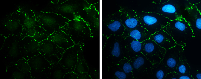
N-Cadherin antibody [N1N3] detects N-Cadherin protein at cell membrane by immunofluorescent analysis. Sample: NT2D1 cells were fixed in 4% paraformaldehyde at RT for 15 min. Green: N-Cadherin stained by N-Cadherin antibody [N1N3] (GTX112734) diluted at 1:500. Blue: Hoechst 33342 staining.
![Various whole cell extracts (30 μg) were separated by 5% SDS-PAGE, and the membrane was blotted with N-Cadherin antibody [N1N3] (GTX112734) diluted at 1:1000. The HRP-conjugated anti-rabbit IgG antibody (GTX213110-01) was used to detect the primary antibody.](https://www.genetex.com/upload/website/prouct_img/normal/GTX112734/GTX112734_42957_20170908_WB_w_23060500_381.webp "Various whole cell extracts (30 μg) were separated by 5% SDS-PAGE, and the membrane was blotted with N-Cadherin antibody [N1N3] (GTX112734) diluted at 1:1000. The HRP-conjugated anti-rabbit IgG antibody (GTX213110-01) was used to detect the primary antibody.")
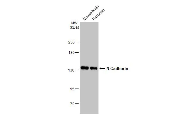
![Various tissue extracts (50 μg) were separated by 5% SDS-PAGE, and the membrane was blotted with N-Cadherin antibody [N1N3] (GTX112734) diluted at 1:1000. The HRP-conjugated anti-rabbit IgG antibody (GTX213110-01) was used to detect the primary antibody.](https://www.genetex.com/upload/website/prouct_img/normal/GTX112734/GTX112734_42957_20170915_WB_M_R_w_23060500_157.webp "Various tissue extracts (50 μg) were separated by 5% SDS-PAGE, and the membrane was blotted with N-Cadherin antibody [N1N3] (GTX112734) diluted at 1:1000. The HRP-conjugated anti-rabbit IgG antibody (GTX213110-01) was used to detect the primary antibody.")
![N-Cadherin antibody [N1N3] detects N-Cadherin protein by western blot analysis. A. 30 μg PC-12 whole cell extract B. 30 μg Rat2 whole cell extract 5% SDS-PAGE N-Cadherin antibody [N1N3] (GTX112734) dilution: 1:1000 The HRP-conjugated anti-rabbit IgG antibody (GTX213110-01) was used to detect the primary antibody.](https://www.genetex.com/upload/website/prouct_img/normal/GTX112734/GTX112734_40394_WB_R_w_23060500_869.webp "N-Cadherin antibody [N1N3] detects N-Cadherin protein by western blot analysis. A. 30 μg PC-12 whole cell extract B. 30 μg Rat2 whole cell extract 5% SDS-PAGE N-Cadherin antibody [N1N3] (GTX112734) dilution: 1:1000 The HRP-conjugated anti-rabbit IgG antibody (GTX213110-01) was used to detect the primary antibody.")
![N-Cadherin antibody [N1N3] detects N-Cadherin protein at cell membrane by immunohistochemical analysis. Sample: Paraffin-embedded mouse liver. N-Cadherin stained by N-Cadherin antibody [N1N3] (GTX112734) diluted at 1:500. Antigen Retrieval: Citrate buffer, pH 6.0, 15 min](https://www.genetex.com/upload/website/prouct_img/normal/GTX112734/GTX112734_42957_20190628_IHC-P_M_w_23060500_326.webp "N-Cadherin antibody [N1N3] detects N-Cadherin protein at cell membrane by immunohistochemical analysis. Sample: Paraffin-embedded mouse liver. N-Cadherin stained by N-Cadherin antibody [N1N3] (GTX112734) diluted at 1:500. Antigen Retrieval: Citrate buffer, pH 6.0, 15 min")
 was separated by 7.5% SDS-PAGE, and the membrane was blotted with N-cadherin antibody (GTX112734) diluted at 1:1000. The HRP-conjugated anti-rabbit IgG antibody (GTX213110-01) was used to detect the primary antibody.")
![N-Cadherin antibody [N1N3] detects CDH2 protein at membrane on hepatoma by immunohistochemical analysis. Sample: Paraffin-embedded human hepatoma. N-Cadherin antibody [N1N3] (GTX112734) dilution: 1:500.
Antigen Retrieval: Trilogy? (EDTA based, pH 8.0) buffer, 15min](https://www.genetex.com/upload/website/prouct_img/normal/GTX112734/GTX112734_40394_IHC_2_w_23060500_268.webp "N-Cadherin antibody [N1N3] detects CDH2 protein at membrane on hepatoma by immunohistochemical analysis. Sample: Paraffin-embedded human hepatoma. N-Cadherin antibody [N1N3] (GTX112734) dilution: 1:500.
Antigen Retrieval: Trilogy? (EDTA based, pH 8.0) buffer, 15min")
![N-Cadherin antibody [N1N3] detects N-Cadherin protein at cell membrane by immunohistochemical analysis. Sample: Paraffin-embedded rat liver. N-Cadherin stained by N-Cadherin antibody [N1N3] (GTX112734) diluted at 1:500. Antigen Retrieval: Citrate buffer, pH 6.0, 15 min](https://www.genetex.com/upload/website/prouct_img/normal/GTX112734/GTX112734_42957_20190628_IHC-P_R_w_23060500_844.webp "N-Cadherin antibody [N1N3] detects N-Cadherin protein at cell membrane by immunohistochemical analysis. Sample: Paraffin-embedded rat liver. N-Cadherin stained by N-Cadherin antibody [N1N3] (GTX112734) diluted at 1:500. Antigen Retrieval: Citrate buffer, pH 6.0, 15 min")
![Wild-type (WT) and N-Cadherin knockout (KO) 293T cell extracts (30 μg) were separated by 5% SDS-PAGE, and the membrane was blotted with N-Cadherin antibody [N1N3] (GTX112734) diluted at 1:500. The HRP-conjugated anti-rabbit IgG antibody (GTX213110-01) was used to detect the primary antibody.](https://www.genetex.com/upload/website/prouct_img/normal/GTX112734/GTX112734_42957_20180810_WB_KO_watermark_w_23060500_824.webp "Wild-type (WT) and N-Cadherin knockout (KO) 293T cell extracts (30 μg) were separated by 5% SDS-PAGE, and the membrane was blotted with N-Cadherin antibody [N1N3] (GTX112734) diluted at 1:500. The HRP-conjugated anti-rabbit IgG antibody (GTX213110-01) was used to detect the primary antibody.")
![Whole cell extract (30 μg) was separated by 7.5% SDS-PAGE, and the membrane was blotted with N-Cadherin antibody [N1N3] (GTX112734) diluted at 1:1000. The HRP-conjugated anti-rabbit IgG antibody (GTX213110-01) was used to detect the primary antibody.](https://www.genetex.com/upload/website/prouct_img/normal/GTX112734/GTX112734_40394_20180316_WB_w_23060500_529.webp "Whole cell extract (30 μg) was separated by 7.5% SDS-PAGE, and the membrane was blotted with N-Cadherin antibody [N1N3] (GTX112734) diluted at 1:1000. The HRP-conjugated anti-rabbit IgG antibody (GTX213110-01) was used to detect the primary antibody.")
N-Cadherin antibody [N1N3] detects N-Cadherin protein at cell membrane by immunofluorescent analysis. Sample: NT2D1 cells were fixed in 4% paraformaldehyde at RT for 15 min. Green: N-Cadherin stained by N-Cadherin antibody [N1N3] (GTX112734) diluted at 1:500. Blue: Hoechst 33342 staining.
N-Cadherin antibody [N1N3]
GTX112734
ApplicationsImmunoFluorescence, Western Blot, ImmunoCytoChemistry, ImmunoHistoChemistry, ImmunoHistoChemistry Paraffin
Product group Antibodies
ReactivityHuman, Mouse, Rat
TargetCDH2
Overview
- SupplierGeneTex
- Product NameN-Cadherin antibody [N1N3]
- Delivery Days Customer9
- Application Supplier NoteWB: 1:500-1:3000. ICC/IF: 1:100-1:1000. IHC-P: 1:100-1:1000. *Optimal dilutions/concentrations should be determined by the researcher.Not tested in other applications.
- ApplicationsImmunoFluorescence, Western Blot, ImmunoCytoChemistry, ImmunoHistoChemistry, ImmunoHistoChemistry Paraffin
- CertificationResearch Use Only
- ClonalityPolyclonal
- Concentration0.19 mg/ml
- ConjugateUnconjugated
- Gene ID1000
- Target nameCDH2
- Target descriptioncadherin 2
- Target synonymsACOGS, ADHD8, ARVD14, CD325, CDHN, CDw325, NCAD, cadherin-2, N-cadherin 1, cadherin 2, type 1, N-cadherin (neuronal), calcium-dependent adhesion protein, neuronal, neural cadherin
- HostRabbit
- IsotypeIgG
- Protein IDP19022
- Protein NameCadherin-2
- Scientific DescriptionThis gene is a classical cadherin from the cadherin superfamily. The encoded protein is a calcium dependent cell-cell adhesion glycoprotein comprised of five extracellular cadherin repeats, a transmembrane region and a highly conserved cytoplasmic tail. The protein functions during gastrulation and is required for establishment of left-right asymmetry. At certain central nervous system synapses, presynaptic to postsynaptic adhesion is mediated at least in part by this gene product. [provided by RefSeq]
- ReactivityHuman, Mouse, Rat
- Storage Instruction-20°C or -80°C,2°C to 8°C
- UNSPSC12352203
References
- Zhang MH, Liu J. Cleavage stimulation factor 2 promotes malignant progression of liver hepatocellular carcinoma by activating phosphatidylinositol 3'-kinase/protein kinase B/mammalian target of rapamycin pathway. Bioengineered. 2022,13(4):10047-10060. doi: 10.1080/21655979.2022.2063100Read this paper
- Chang YX, Lin YF, Chen CL, et al. Chaperonin-Containing TCP-1 Promotes Cancer Chemoresistance and Metastasis through the AKT-GSK3β-β-Catenin and XIAP-Survivin Pathways. Cancers (Basel). 2020,12(12). doi: 10.3390/cancers12123865Read this paper
- Ho KH, Cheng CH, Chou CM, et al. miR-140 targeting CTSB signaling suppresses the mesenchymal transition and enhances temozolomide cytotoxicity in glioblastoma multiforme. Pharmacol Res. 2019,147:104390. doi: 10.1016/j.phrs.2019.104390Read this paper
- Ling HH, Kuo CC, Lin BX, et al. Elevation of YAP promotes the epithelial-mesenchymal transition and tumor aggressiveness in colorectal cancer. Exp Cell Res. 2017,350(1):218-225. doi: 10.1016/j.yexcr.2016.11.024Read this paper
- Chung IH, Wu TI, Liao CJ, et al. Overexpression of lipocalin 2 in human cervical cancer enhances tumor invasion. Oncotarget. 2016,7(10):11113-26. doi: 10.18632/oncotarget.7096Read this paper
- Jian MY, Liu Y, Li Q, et al. N-cadherin coordinates AMP kinase-mediated lung vascular repair. Am J Physiol Lung Cell Mol Physiol. 2016,310(1):L71-85. doi: 10.1152/ajplung.00227.2015Read this paper
- Hsu TH, Jiang SY, Chang WL, et al. Involvement of RARRES3 in the regulation of Wnt proteins acylation and signaling activities in human breast cancer cells. Cell Death Differ. 2015,22(5):801-14. doi: 10.1038/cdd.2014.175Read this paper
- Lupia A, Peppicelli S, Witort E, et al. CD63 tetraspanin is a negative driver of epithelial-to-mesenchymal transition in human melanoma cells. J Invest Dermatol. 2014,134(12):2947-2956. doi: 10.1038/jid.2014.258Read this paper
Datasheet
Related products
Product group Antibodies

Anti-N-cadherin [8C11]AB01660-1.1-BT
ApplicationsFlow Cytometry, ImmunoFluorescence, Western Blot, ImmunoHistoChemistry
ReactivityHamster, Human, Rabbit
TargetCDH2
- SizePrice
Product group Antibodies
Anti-CDH2 Antibody144-03045
ApplicationsImmunoPrecipitation, Western Blot
ReactivityHuman, Mouse, Rat
TargetCDH2
- SizePrice

Product group Antibodies
Anti-CDH2 AntibodyAMAB91220
ApplicationsWestern Blot, ImmunoCytoChemistry, ImmunoHistoChemistry
ReactivityHuman
TargetCDH2
- SizePrice
Product group Antibodies

N-Cadherin AntibodyABX123447
ApplicationsWestern Blot, ImmunoHistoChemistry
- SizePrice

Product group Antibodies
Anti-N-Cadherin-2 CDH2 CD325-Antibody Picoband(r)A01577-2-CARRIER-FREE
ApplicationsWestern Blot
ReactivityHuman
TargetCDH2
- SizePrice
Product group Antibodies
References
N-Cadherin antibodyGTX127345
ApplicationsImmunoFluorescence, Western Blot, ImmunoCytoChemistry, ImmunoHistoChemistry, ImmunoHistoChemistry Frozen, ImmunoHistoChemistry Paraffin
ReactivityHuman, Monkey, Mouse, Rat
TargetCDH2
- SizePrice
![Wild-type (WT) and N-Cadherin knockout (KO) 293T cell extracts (30 μg) were separated by 5% SDS-PAGE, and the membrane was blotted with N-Cadherin antibody [N2C1], Internal (GTX112733) diluted at 1:500. The HRP-conjugated anti-rabbit IgG antibody (GTX213110-01) was used to detect the primary antibody.](https://www.genetex.com/upload/website/prouct_img/normal/GTX112733/GTX112733_40394_20180810_WB_KO_watermark_w_23060500_602.webp)
Product group Antibodies
References
N-Cadherin antibody [N2C1], InternalGTX112733
ApplicationsImmunoFluorescence, Western Blot, ImmunoCytoChemistry, ImmunoHistoChemistry, ImmunoHistoChemistry Frozen, ImmunoHistoChemistry Paraffin
ReactivityHuman, Mouse, Rat
TargetCDH2
- SizePrice
![IHC-P analysis of human lung cancer (A), colon cancer (B), ovarian cancer (C) and mammary cancer(D) using GTX82992 N-Cadherin antibody [5D5].](https://www.genetex.com/upload/website/prouct_img/normal/GTX82992/GTX82992_20170912_IHC-P_w_23061322_220.webp)
Product group Antibodies
References
N-Cadherin antibody [5D5]GTX82992
ApplicationsFlow Cytometry, ImmunoFluorescence, Western Blot, ELISA, ImmunoCytoChemistry, ImmunoHistoChemistry, ImmunoHistoChemistry Paraffin
ReactivityHuman, Mouse, Rat
TargetCDH2
- SizePrice
Product group Antibodies
CDH2 Polyclonal AntibodyCAC14634
ApplicationsImmunoFluorescence, Western Blot, ELISA
ReactivityMouse
TargetCDH2
- SizePrice