

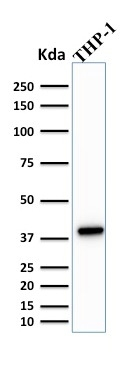
WB analysis of THP-1 lysate using GTX17997 PU.1 antibody [PU1/2146].
![IHC-P analysis of human lymph node tissue using GTX17997 PU.1 antibody [PU1/2146].](https://www.genetex.com/upload/website/prouct_img/normal/GTX17997/GTX17997_20200115_IHC-P_717_w_23060620_868.webp "IHC-P analysis of human lymph node tissue using GTX17997 PU.1 antibody [PU1/2146].")
. Z- and S- Score: The Z-score represents the strength of a signal that a monoclonal antibody produces when binding to a particular protein on the HuProtTM array. Z-scores are described in units of standard deviations (SD’s) above the mean value of all signals generated on that array. If targets on HuProtTM are arranged in descending order of the Z-score, the S-score is the difference (also in units of SD’s) between the Z-score. S-score therefore represents the relative target specificity of a Monoclonal Antibody to its intended target. A Monoclonal Antibody is considered to specific to its intended target if the Monoclonal Antibody has an S-score of at least 2.5. For example, if a Monoclonal Antibody binds to protein X with a Z-score of 43 and to protein Y with a Z-score of 14, then the S-score for the binding of that Monoclonal Antibody to protein X is equal to 29.")
![IHC-P analysis of human colon carcinoma tissue using GTX17997 PU.1 antibody [PU1/2146].](https://www.genetex.com/upload/website/prouct_img/normal/GTX17997/GTX17997_20200115_IHC-P_499_w_23060620_545.webp "IHC-P analysis of human colon carcinoma tissue using GTX17997 PU.1 antibody [PU1/2146].")
![IHC-P analysis of human spleen tissue using GTX17997 PU.1 antibody [PU1/2146].](https://www.genetex.com/upload/website/prouct_img/normal/GTX17997/GTX17997_20200115_IHC-P_1170_w_23060620_491.webp "IHC-P analysis of human spleen tissue using GTX17997 PU.1 antibody [PU1/2146].")
![FACS analysis of PFA-fixed Ramos cells using GTX17997 PU.1 antibody [PU1/2146]. Blue : Primary antibody Red : Isotype control](https://www.genetex.com/upload/website/prouct_img/normal/GTX17997/GTX17997_20200115_FACS_1755_w_23060620_945.webp "FACS analysis of PFA-fixed Ramos cells using GTX17997 PU.1 antibody [PU1/2146]. Blue : Primary antibody Red : Isotype control")
![ICC/IF analysis of PFA-fixed Ramos cells using GTX17997 PU.1 antibody [PU1/2146]. Green : Primary antibody Red : Phalloidin](https://www.genetex.com/upload/website/prouct_img/normal/GTX17997/GTX17997_20200115_ICC-IF_1756_w_23060620_971.webp "ICC/IF analysis of PFA-fixed Ramos cells using GTX17997 PU.1 antibody [PU1/2146]. Green : Primary antibody Red : Phalloidin")
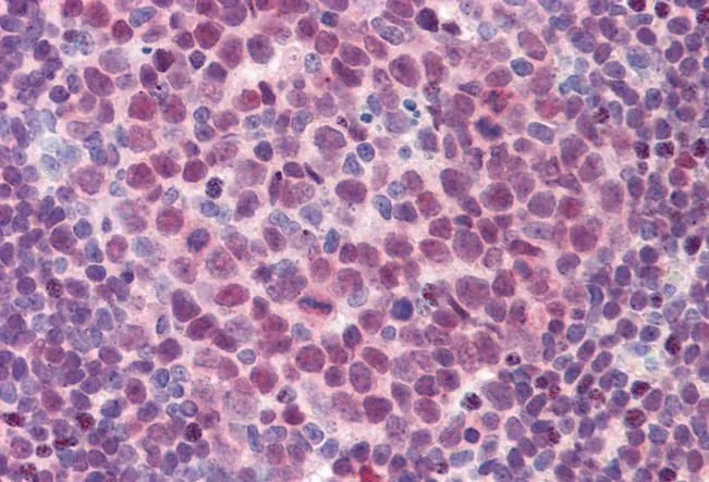
![IHC-P analysis of human Hodgkin's lymphoma tissue using GTX17997 PU.1 antibody [PU1/2146].](https://www.genetex.com/upload/website/prouct_img/normal/GTX17997/GTX17997_20200115_IHC-P_646_w_23060620_208.webp "IHC-P analysis of human Hodgkin's lymphoma tissue using GTX17997 PU.1 antibody [PU1/2146].")
WB analysis of THP-1 lysate using GTX17997 PU.1 antibody [PU1/2146].
PU.1 antibody [PU1/2146]
GTX17997
ApplicationsFlow Cytometry, ImmunoFluorescence, Western Blot, ImmunoCytoChemistry, ImmunoHistoChemistry, ImmunoHistoChemistry Paraffin, Other Application
Product group Antibodies
ReactivityHuman
TargetSPI1
Overview
- SupplierGeneTex
- Product NamePU.1 antibody [PU1/2146]
- Delivery Days Customer9
- Application Supplier NoteWB: 1-2microg/ml. ICC/IF: 1-2microg/ml. IHC-P: 1-2microg/ml for 30 minutes at RT. FCM: 1-2microg/106 cells. *Optimal dilutions/concentrations should be determined by the researcher.Not tested in other applications.
- ApplicationsFlow Cytometry, ImmunoFluorescence, Western Blot, ImmunoCytoChemistry, ImmunoHistoChemistry, ImmunoHistoChemistry Paraffin, Other Application
- CertificationResearch Use Only
- ClonalityMonoclonal
- Clone IDPU1/2146
- Concentration0.2 mg/ml
- ConjugateUnconjugated
- Gene ID6688
- Target nameSPI1
- Target descriptionSpi-1 proto-oncogene
- Target synonymsAGM10, OF, PU.1, SFPI1, SPI-1, SPI-A, transcription factor PU.1, 31 kDa transforming protein, hematopoietic transcription factor PU.1, spleen focus forming virus (SFFV) proviral integration oncogene spi1
- HostMouse
- IsotypeIgG2b
- Protein IDP17947
- Protein NameTranscription factor PU.1
- Scientific DescriptionThis gene encodes an ETS-domain transcription factor that activates gene expression during myeloid and B-lymphoid cell development. The nuclear protein binds to a purine-rich sequence known as the PU-box found near the promoters of target genes, and regulates their expression in coordination with other transcription factors and cofactors. The protein can also regulate alternative splicing of target genes. Multiple transcript variants encoding different isoforms have been found for this gene. [provided by RefSeq, Jul 2008]
- ReactivityHuman
- Storage Instruction-20°C or -80°C,2°C to 8°C
- UNSPSC41116161
Datasheet
Related products

Product group Antibodies
Anti-SPI1 AntibodyA97901
ApplicationsWestern Blot, ELISA
ReactivityHuman, Mouse, Rat
- SizePrice

Product group Antibodies
Anti-PU.1/SPI1 Antibody Picoband(r)A01116-1-CARRIER-FREE
ApplicationsFlow Cytometry, Western Blot, ELISA, ImmunoHistoChemistry
ReactivityHuman, Mouse, Rat
TargetSPI1
- SizePrice
Product group Antibodies

SPI1 / PU.1 AntibodyLS-C831700
ApplicationsWestern Blot, ELISA
ReactivityHuman, Mouse, Rat
TargetSPI1
- SizePrice
Product group Antibodies
PU.1/Spi1 Polyclonal AntibodyBS-19594R
ApplicationsImmunoFluorescence, Western Blot, ELISA, ImmunoCytoChemistry, ImmunoHistoChemistry, ImmunoHistoChemistry Frozen, ImmunoHistoChemistry Paraffin
ReactivityBovine, Equine, Human, Mouse, Porcine, Rat, Sheep
TargetSPI1
- SizePrice
Product group Antibodies
SPI1 AntibodyCSB-PA003887
ApplicationsImmunoFluorescence, Western Blot, ELISA, ImmunoHistoChemistry
ReactivityHuman, Monkey, Mouse, Rat
TargetSPI1
- SizePrice
Product group Antibodies

Goat anti-PU.1EB08429
ApplicationsWestern Blot, ELISA, ImmunoHistoChemistry
ReactivityHuman, Rat
TargetSPI1
- SizePrice
![THP-1 whole cell and nuclear extracts (30 μg) were separated by 10% SDS-PAGE, and the membrane was blotted with PU.1 antibody [N1C1] (GTX101581) diluted at 1:500. The HRP-conjugated anti-rabbit IgG antibody (GTX213110-01) was used to detect the primary antibody.](https://www.genetex.com/upload/website/prouct_img/normal/GTX101581/GTX101581_43600_20190607_WB_Fraction_w_23060100_464.webp)
Product group Antibodies
PU.1 antibody [N1C1]GTX101581
ApplicationsWestern Blot
ReactivityHuman, Mouse
TargetSPI1
- SizePrice
Product group Antibodies
PU.1 antibody, InternalGTX88620
ApplicationsWestern Blot, ImmunoHistoChemistry, ImmunoHistoChemistry Paraffin
ReactivityHuman
TargetSPI1
- SizePrice

Product group Antibodies
Anti-SPI1 AntibodyHPA044653
ApplicationsChIP Chromatin ImmunoPrecipitation, ImmunoCytoChemistry, ImmunoHistoChemistry
ReactivityHuman
TargetSPI1
- SizePrice