

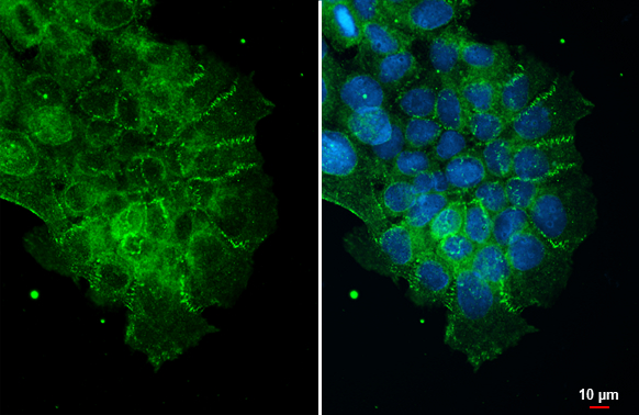
ZO-1 antibody [N1N2], N-term detects ZO-1 protein at cell membrane by immunofluorescent analysis. Sample: A431 cells were fixed in ice-cold MeOH for 5 min. Green: ZO-1 stained by ZO-1 antibody [N1N2], N-term (GTX108613) diluted at 1:500.
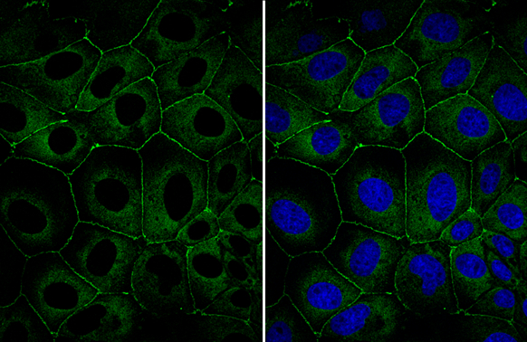
![ZO-1 antibody [N1N2], N-term detects ZO-1 protein at cell membrane by immunofluorescent analysis. Sample: NT2D1 cells were fixed in ice-cold MeOH for 5 min. Green: ZO-1 stained by ZO-1 antibody [N1N2], N-term (GTX108613) diluted at 1:200. Blue: Hoechst 33342 staining.](https://www.genetex.com/upload/website/prouct_img/normal/GTX108613/GTX108613_42753_20171213_ICC_IF_w_23060120_132.webp "ZO-1 antibody [N1N2], N-term detects ZO-1 protein at cell membrane by immunofluorescent analysis. Sample: NT2D1 cells were fixed in ice-cold MeOH for 5 min. Green: ZO-1 stained by ZO-1 antibody [N1N2], N-term (GTX108613) diluted at 1:200. Blue: Hoechst 33342 staining.")
![Non-transfected (–) and transfected (+) HeLa whole cell extracts (30 μg) were separated by 5% SDS-PAGE, and the membrane was blotted with ZO-1 antibody [N1N2], N-term (GTX108613) diluted at 1:1000. The HRP-conjugated anti-rabbit IgG antibody (GTX213110-01) was used to detect the primary antibody.](https://www.genetex.com/upload/website/prouct_img/normal/GTX108613/GTX108613_39967_20160804_WB_shRNA_watermark_w_23060120_586.webp "Non-transfected (–) and transfected (+) HeLa whole cell extracts (30 μg) were separated by 5% SDS-PAGE, and the membrane was blotted with ZO-1 antibody [N1N2], N-term (GTX108613) diluted at 1:1000. The HRP-conjugated anti-rabbit IgG antibody (GTX213110-01) was used to detect the primary antibody.")
![Mouse tissue extract (50 μg) was separated by 5% SDS-PAGE, and the membrane was blotted with ZO-1 antibody [N1N2], N-term (GTX108613) diluted at 1:500. The HRP-conjugated anti-rabbit IgG antibody (GTX213110-01) was used to detect the primary antibody.](https://www.genetex.com/upload/website/prouct_img/normal/GTX108613/GTX108613_43607_20191114_WB_M_testis_w_23060120_838.webp "Mouse tissue extract (50 μg) was separated by 5% SDS-PAGE, and the membrane was blotted with ZO-1 antibody [N1N2], N-term (GTX108613) diluted at 1:500. The HRP-conjugated anti-rabbit IgG antibody (GTX213110-01) was used to detect the primary antibody.")


![Various whole cell extracts (30 μg) were separated by 5% SDS-PAGE, and the membrane was blotted with ZO-1 antibody [N1N2], N-term (GTX108613) diluted at 1:1000. The HRP-conjugated anti-rabbit IgG antibody (GTX213110-01) was used to detect the primary antibody.](https://www.genetex.com/upload/website/prouct_img/normal/GTX108613/GTX108613_43887_20240524_WB_R_24061301_205.webp "Various whole cell extracts (30 μg) were separated by 5% SDS-PAGE, and the membrane was blotted with ZO-1 antibody [N1N2], N-term (GTX108613) diluted at 1:1000. The HRP-conjugated anti-rabbit IgG antibody (GTX213110-01) was used to detect the primary antibody.")
![Various whole cell extracts (30 μg) were separated by 5% SDS-PAGE, and the membrane was blotted with ZO-1 antibody [N1N2], N-term (GTX108613) diluted at 1:500. The HRP-conjugated anti-rabbit IgG antibody (GTX213110-01) was used to detect the primary antibody.](https://www.genetex.com/upload/website/prouct_img/normal/GTX108613/GTX108613_43915_20211119_WB_M_24122400_725.webp "Various whole cell extracts (30 μg) were separated by 5% SDS-PAGE, and the membrane was blotted with ZO-1 antibody [N1N2], N-term (GTX108613) diluted at 1:500. The HRP-conjugated anti-rabbit IgG antibody (GTX213110-01) was used to detect the primary antibody.")
![Various whole cell extracts (30 μg) were separated by 5% SDS-PAGE, and the membrane was blotted with ZO-1 antibody [N1N2], N-term (GTX108613) diluted at 1:1000. The HRP-conjugated anti-rabbit IgG antibody (GTX213110-01) was used to detect the primary antibody.](https://www.genetex.com/upload/website/prouct_img/normal/GTX108613/GTX108613_43915_20200417_WB_24122400_779.webp "Various whole cell extracts (30 μg) were separated by 5% SDS-PAGE, and the membrane was blotted with ZO-1 antibody [N1N2], N-term (GTX108613) diluted at 1:1000. The HRP-conjugated anti-rabbit IgG antibody (GTX213110-01) was used to detect the primary antibody.")
![Various whole cell extracts (30 μg) were separated by 5% SDS-PAGE, and the membrane was blotted with ZO-1 antibody [N1N2], N-term (GTX108613) diluted at 1:500. The HRP-conjugated anti-rabbit IgG antibody (GTX213110-01) was used to detect the primary antibody.](https://www.genetex.com/upload/website/prouct_img/normal/GTX108613/GTX108613_43915_20210827_WB_M_24122400_483.webp "Various whole cell extracts (30 μg) were separated by 5% SDS-PAGE, and the membrane was blotted with ZO-1 antibody [N1N2], N-term (GTX108613) diluted at 1:500. The HRP-conjugated anti-rabbit IgG antibody (GTX213110-01) was used to detect the primary antibody.")
ZO-1 antibody [N1N2], N-term detects ZO-1 protein at cell membrane by immunofluorescent analysis. Sample: A431 cells were fixed in ice-cold MeOH for 5 min. Green: ZO-1 stained by ZO-1 antibody [N1N2], N-term (GTX108613) diluted at 1:500.
ZO-1 antibody [N1N2], N-term
GTX108613
ApplicationsImmunoFluorescence, ImmunoPrecipitation, Western Blot, ImmunoCytoChemistry, ImmunoHistoChemistry, ImmunoHistoChemistry Frozen
Product group Antibodies
ReactivityFish, Human, Mouse, Rat
TargetTJP1
Overview
- SupplierGeneTex
- Product NameZO-1 antibody [N1N2], N-term
- Delivery Days Customer9
- Application Supplier NoteWB: 1:500-1:3000. ICC/IF: 1:100-1:1000. *Optimal dilutions/concentrations should be determined by the researcher.Not tested in other applications.
- ApplicationsImmunoFluorescence, ImmunoPrecipitation, Western Blot, ImmunoCytoChemistry, ImmunoHistoChemistry, ImmunoHistoChemistry Frozen
- CertificationResearch Use Only
- ClonalityPolyclonal
- Concentration0.58 mg/ml
- ConjugateUnconjugated
- Gene ID7082
- Target nameTJP1
- Target descriptiontight junction protein 1
- Target synonymsZO-1, tight junction protein 1, tight junction protein ZO-1, zona occludens 1, zonula occludens 1 protein
- HostRabbit
- IsotypeIgG
- Protein IDQ07157
- Protein NameTight junction protein 1
- Scientific DescriptionThis gene encodes a protein located on a cytoplasmic membrane surface of intercellular tight junctions. The encoded protein may be involved in signal transduction at cell-cell junctions. Two transcript variants encoding distinct isoforms have been identified for this gene. [provided by RefSeq]
- ReactivityFish, Human, Mouse, Rat
- Storage Instruction-20°C or -80°C,2°C to 8°C
- UNSPSC12352203
References
- Hsia Y, Sivasubramanian M, Chu CH, et al. A Dual Concentration-Tailored Cytokine-Chemo Nanosystem to Alleviate Multidrug Resistance and Redirect Balance of Cancer Proliferation and Apoptosis. Int J Nanomedicine. 2023,18:4253-4274. doi: 10.2147/IJN.S412932Read this paper
- Su WP, Li CJ, Lin LT, et al. Boosting mitochondrial function and metabolism in aging female germ cells with dual ROCK/ROS inhibition. Biomed Pharmacother. 2023,163:114888. doi: 10.1016/j.biopha.2023.114888Read this paper
- Möckel M, Baldok N, Walles T, et al. Human 3D Airway Tissue Models for Real-Time Microscopy: Visualizing Respiratory Virus Spreading. Cells. 2022,11(22). doi: 10.3390/cells11223634Read this paper
- Russell DF, Zhang W, Warnock TC, et al. Lectin binding and gel secretion within Lorenzinian electroreceptors of Polyodon. PLoS One. 2022,17(11):e0276854. doi: 10.1371/journal.pone.0276854Read this paper
- Odom MR, Hughes FM Jr, Jin H, et al. Diabetes causes NLRP3-dependent barrier dysfunction in mice with detrusor overactivity but not underactivity. Am J Physiol Renal Physiol. 2022,323(6):F616-F632. doi: 10.1152/ajprenal.00047.2022Read this paper
- Zhang MH, Liu J. Cleavage stimulation factor 2 promotes malignant progression of liver hepatocellular carcinoma by activating phosphatidylinositol 3'-kinase/protein kinase B/mammalian target of rapamycin pathway. Bioengineered. 2022,13(4):10047-10060. doi: 10.1080/21655979.2022.2063100Read this paper
- Liu X, Chen B, Chen J, et al. Deubiquitinase ubiquitin-specific peptidase 10 maintains cysteine rich angiogenic inducer 61 expression via Yes1 associated transcriptional regulator to augment immune escape and metastasis of pancreatic adenocarcinoma. Cancer Sci. 2022,113(5):1868-1879. doi: 10.1111/cas.15326Read this paper
- Yen YT, Yang JC, Chang JB, et al. Down-Regulation of miR-194-5p for Predicting Metastasis in Breast Cancer Cells. Int J Mol Sci. 2021,23(1). doi: 10.3390/ijms23010325Read this paper
- Russell DF, Warnock TC, Zhang W, et al. Large-Scale Convergence of Receptor Cell Arrays Onto Afferent Terminal Arbors in the Lorenzinian Electroreceptors of Polyodon. Front Neuroanat. 2020,14:50. doi: 10.3389/fnana.2020.00050Read this paper
- Voirin AC, Celle S, Perek N, et al. Sera of elderly obstructive sleep apnea patients alter blood-brain barrier integrity in vitro: a pilot study. Sci Rep. 2020,10(1):11309. doi: 10.1038/s41598-020-68374-8Read this paper
Datasheet
Related products
Product group Antibodies
Anti-ZO-1 Antibody144-61634
ApplicationsImmunoFluorescence, Western Blot
ReactivityHuman, Mouse, Rat
TargetTJP1
- SizePrice
![Non-transfected (–) and transfected (+) HeLa whole cell extracts (30 μg) were separated by 5% SDS-PAGE, and the membrane was blotted with ZO-1 antibody [C3], C-term (GTX108587) diluted at 1:500.](https://www.genetex.com/upload/website/prouct_img/normal/GTX108587/GTX108587_40618_20160804_WB_shRNA_watermark_w_23060120_565.webp)
Product group Antibodies
References
ZO-1 antibody [C3], C-termGTX108587
ApplicationsWestern Blot
ReactivityHuman
TargetTJP1
- SizePrice
Product group Antibodies
References
ZO-1 antibodyGTX108592
ApplicationsImmunoFluorescence, Western Blot, ImmunoCytoChemistry, ImmunoHistoChemistry, ImmunoHistoChemistry Frozen, ImmunoHistoChemistry Paraffin
ReactivityBovine, Canine, Human, Monkey, Mouse, Porcine
TargetTJP1
- SizePrice
![Non-transfected (–) and transfected (+) HeLa whole cell extracts (30 μg) were separated by 5% SDS-PAGE, and the membrane was blotted with ZO-1 antibody [N2C1], Internal (GTX108627) diluted at 1:500. The HRP-conjugated anti-rabbit IgG antibody (GTX213110-01) was used to detect the primary antibody.](https://www.genetex.com/upload/website/prouct_img/normal/GTX108627/GTX108627_40464_20160804_WB_shRNA_watermark_w_23060120_175.webp)
Product group Antibodies
References
ZO-1 antibody [N2C1], InternalGTX108627
ApplicationsImmunoFluorescence, Western Blot, ImmunoCytoChemistry, ImmunoHistoChemistry, ImmunoHistoChemistry Frozen
ReactivityHuman, Rat
TargetTJP1
- SizePrice
![Wild-type (WT) and TJP1 knockout (KO) HeLa cell extracts (30 μg) were separated by 5% SDS-PAGE, and the membrane was blotted with ZO-1 antibody [HL1133] (GTX636399) diluted at 1:1000. The HRP-conjugated anti-rabbit IgG antibody (GTX213110-01) was used to detect the primary antibody.](https://www.genetex.com/upload/website/prouct_img/normal/GTX636399/GTX636399_44466_20220211_WB_KO_watermark_w_23061202_165.webp)
Product group Antibodies
References
ZO-1 antibody [HL1133]GTX636399
ApplicationsImmunoFluorescence, Western Blot, ImmunoCytoChemistry
ReactivityHuman
TargetTJP1
- SizePrice
![ZO-1 antibody [HL1185] detects ZO-1 protein at cell membrane by immunofluorescent analysis. Sample: A431 cells were fixed in 4% paraformaldehyde at RT for 15 min. Green: ZO-1 stained by ZO-1 antibody [HL1185] (GTX636491) diluted at 1:500. Blue: Fluoroshield with DAPI (GTX30920).](https://www.genetex.com/upload/website/prouct_img/normal/GTX636491/GTX636491_T-44445_20211126_ICC_IF_w_23061202_412.webp)
Product group Antibodies
ZO-1 antibody [HL1185]GTX636491
ApplicationsImmunoFluorescence, Western Blot, ImmunoCytoChemistry
ReactivityCanine, Feline, Human, Mouse, Rat
TargetTJP1
- SizePrice
Product group Antibodies
Tjp1 Polyclonal AntibodyCAC11695
ApplicationsImmunoFluorescence, ELISA
TargetTJP1
- SizePrice
Product group Antibodies
References
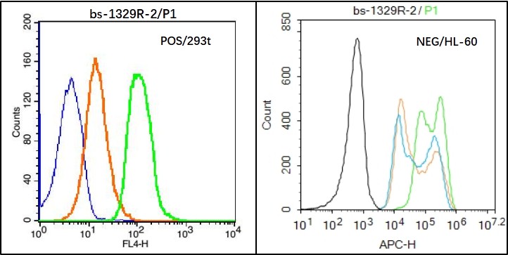
ZO-1/TJP1 Polyclonal AntibodyBS-1329R
ApplicationsFlow Cytometry, ImmunoFluorescence, Western Blot, ELISA, ImmunoCytoChemistry, ImmunoHistoChemistry, ImmunoHistoChemistry Frozen, ImmunoHistoChemistry Paraffin
ReactivityBovine, Canine, Chicken, Guinea Pig, Goat, Human, Mouse, Porcine, Rat, Sheep
TargetTJP1
- SizePrice