

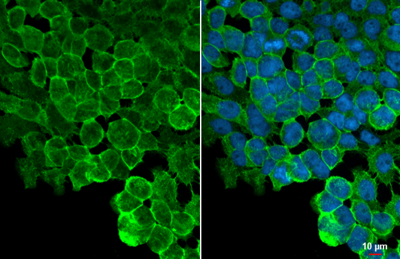
ZO-1 antibody [HL1185] detects ZO-1 protein at cell membrane by immunofluorescent analysis. Sample: A431 cells were fixed in 4% paraformaldehyde at RT for 15 min. Green: ZO-1 stained by ZO-1 antibody [HL1185] (GTX636491) diluted at 1:500. Blue: Fluoroshield with DAPI (GTX30920).
![Various whole cell extracts (30 μg) were separated by 5% SDS-PAGE, and the membrane was blotted with ZO-1 antibody [HL1185] (GTX636491) diluted at 1:1000. The HRP-conjugated anti-rabbit IgG antibody (GTX213110-01) was used to detect the primary antibody.](https://www.genetex.com/upload/website/prouct_img/normal/GTX636491/GTX636491_T-44445_20220107_WB_M_w_23061202_894.webp "Various whole cell extracts (30 μg) were separated by 5% SDS-PAGE, and the membrane was blotted with ZO-1 antibody [HL1185] (GTX636491) diluted at 1:1000. The HRP-conjugated anti-rabbit IgG antibody (GTX213110-01) was used to detect the primary antibody.")
![Wild-type (WT) and TJP1 knockout (KO) HeLa cell extracts (30 μg) were separated by 5% SDS-PAGE, and the membrane was blotted with ZO-1 antibody [HL1185] (GTX636491) diluted at 1:1000. The HRP-conjugated anti-rabbit IgG antibody (GTX213110-01) was used to detect the primary antibody.](https://www.genetex.com/upload/website/prouct_img/normal/GTX636491/GTX636491_44550_20220211_WB_KO_watermark_w_23061202_583.webp "Wild-type (WT) and TJP1 knockout (KO) HeLa cell extracts (30 μg) were separated by 5% SDS-PAGE, and the membrane was blotted with ZO-1 antibody [HL1185] (GTX636491) diluted at 1:1000. The HRP-conjugated anti-rabbit IgG antibody (GTX213110-01) was used to detect the primary antibody.")
![Various whole cell extracts (30 μg) were separated by 5% SDS-PAGE, and the membrane was blotted with ZO-1 antibody [HL1185] (GTX636491) diluted at 1:1000. The HRP-conjugated anti-rabbit IgG antibody (GTX213110-01) was used to detect the primary antibody.](https://www.genetex.com/upload/website/prouct_img/normal/GTX636491/GTX636491_44746_20221209_WB_D_C_23071223_684.webp "Various whole cell extracts (30 μg) were separated by 5% SDS-PAGE, and the membrane was blotted with ZO-1 antibody [HL1185] (GTX636491) diluted at 1:1000. The HRP-conjugated anti-rabbit IgG antibody (GTX213110-01) was used to detect the primary antibody.")
![Various whole cell extracts (30 μg) were separated by 5% SDS-PAGE, and the membrane was blotted with ZO-1 antibody [HL1185] (GTX636491) diluted at 1:1000. The HRP-conjugated anti-rabbit IgG antibody (GTX213110-01) was used to detect the primary antibody.](https://www.genetex.com/upload/website/prouct_img/normal/GTX636491/GTX636491_45278_20240524_WB_R_24052802_865.webp "Various whole cell extracts (30 μg) were separated by 5% SDS-PAGE, and the membrane was blotted with ZO-1 antibody [HL1185] (GTX636491) diluted at 1:1000. The HRP-conjugated anti-rabbit IgG antibody (GTX213110-01) was used to detect the primary antibody.")
![Various whole cell extracts (30 μg) were separated by 5% SDS-PAGE, and the membrane was blotted with ZO-1 antibody [HL1133] (GTX636491) diluted at 1:1000. The HRP-conjugated anti-rabbit IgG antibody (GTX213110-01) was used to detect the primary antibody.](https://www.genetex.com/upload/website/prouct_img/normal/GTX636491/GTX636491_45278_20240112_WB_24052802_887.webp "Various whole cell extracts (30 μg) were separated by 5% SDS-PAGE, and the membrane was blotted with ZO-1 antibody [HL1133] (GTX636491) diluted at 1:1000. The HRP-conjugated anti-rabbit IgG antibody (GTX213110-01) was used to detect the primary antibody.")
ZO-1 antibody [HL1185] detects ZO-1 protein at cell membrane by immunofluorescent analysis. Sample: A431 cells were fixed in 4% paraformaldehyde at RT for 15 min. Green: ZO-1 stained by ZO-1 antibody [HL1185] (GTX636491) diluted at 1:500. Blue: Fluoroshield with DAPI (GTX30920).
ZO-1 antibody [HL1185]
GTX636491
ApplicationsImmunoFluorescence, Western Blot, ImmunoCytoChemistry
Product group Antibodies
ReactivityCanine, Feline, Human, Mouse, Rat
TargetTJP1
Overview
- SupplierGeneTex
- Product NameZO-1 antibody [HL1185]
- Delivery Days Customer9
- Application Supplier NoteICC/IF: 1:100-1:1000. *Optimal dilutions/concentrations should be determined by the researcher.Not tested in other applications.
- ApplicationsImmunoFluorescence, Western Blot, ImmunoCytoChemistry
- CertificationResearch Use Only
- ClonalityMonoclonal
- Clone IDHL1185
- Concentration1 mg/ml
- ConjugateUnconjugated
- Gene ID7082
- Target nameTJP1
- Target descriptiontight junction protein 1
- Target synonymsZO-1, tight junction protein 1, tight junction protein ZO-1, zona occludens 1, zonula occludens 1 protein
- HostRabbit
- IsotypeIgG
- Protein IDQ07157
- Protein NameTight junction protein 1
- Scientific DescriptionThis gene encodes a member of the membrane-associated guanylate kinase (MAGUK) family of proteins, and acts as a tight junction adaptor protein that also regulates adherens junctions. Tight junctions regulate the movement of ions and macromolecules between endothelial and epithelial cells. The multidomain structure of this scaffold protein, including a postsynaptic density 95/disc-large/zona occludens (PDZ) domain, a Src homology (SH3) domain, a guanylate kinase (GuK) domain and unique (U) motifs all help to co-ordinate binding of transmembrane proteins, cytosolic proteins, and F-actin, which are required for tight junction function. Alternative splicing results in multiple transcript variants encoding different isoforms. [provided by RefSeq, Aug 2017]
- ReactivityCanine, Feline, Human, Mouse, Rat
- Storage Instruction-20°C or -80°C,2°C to 8°C
- UNSPSC12352203
Datasheet
Related products
Product group Antibodies
Anti-ZO-1 Antibody144-61634
ApplicationsImmunoFluorescence, Western Blot
ReactivityHuman, Mouse, Rat
TargetTJP1
- SizePrice
![Non-transfected (–) and transfected (+) HeLa whole cell extracts (30 μg) were separated by 5% SDS-PAGE, and the membrane was blotted with ZO-1 antibody [C3], C-term (GTX108587) diluted at 1:500.](https://www.genetex.com/upload/website/prouct_img/normal/GTX108587/GTX108587_40618_20160804_WB_shRNA_watermark_w_23060120_565.webp)
Product group Antibodies
References
ZO-1 antibody [C3], C-termGTX108587
ApplicationsWestern Blot
ReactivityHuman
TargetTJP1
- SizePrice
Product group Antibodies
References
ZO-1 antibodyGTX108592
ApplicationsImmunoFluorescence, Western Blot, ImmunoCytoChemistry, ImmunoHistoChemistry, ImmunoHistoChemistry Frozen, ImmunoHistoChemistry Paraffin
ReactivityBovine, Canine, Human, Monkey, Mouse, Porcine
TargetTJP1
- SizePrice
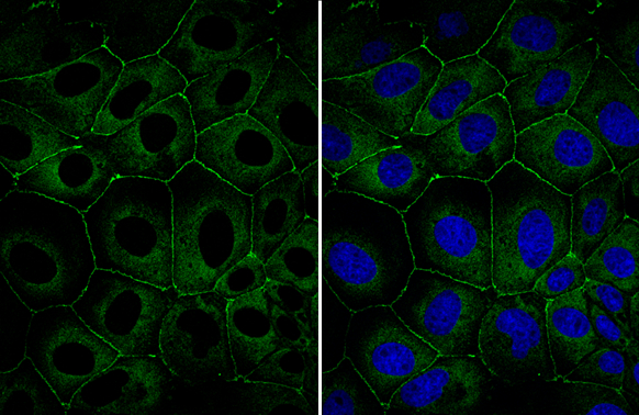
![ZO-1 antibody [N1N2], N-term detects ZO-1 protein at cell membrane by immunofluorescent analysis. Sample: A431 cells were fixed in ice-cold MeOH for 5 min. Green: ZO-1 stained by ZO-1 antibody [N1N2], N-term (GTX108613) diluted at 1:500.](https://www.genetex.com/upload/website/prouct_img/normal/GTX108613/GTX108613_43803_20210416_ICC_IF_w_23060120_527.webp)
Product group Antibodies
References
ZO-1 antibody [N1N2], N-termGTX108613
ApplicationsImmunoFluorescence, ImmunoPrecipitation, Western Blot, ImmunoCytoChemistry, ImmunoHistoChemistry, ImmunoHistoChemistry Frozen
ReactivityFish, Human, Mouse, Rat
TargetTJP1
- SizePrice
![Non-transfected (–) and transfected (+) HeLa whole cell extracts (30 μg) were separated by 5% SDS-PAGE, and the membrane was blotted with ZO-1 antibody [N2C1], Internal (GTX108627) diluted at 1:500. The HRP-conjugated anti-rabbit IgG antibody (GTX213110-01) was used to detect the primary antibody.](https://www.genetex.com/upload/website/prouct_img/normal/GTX108627/GTX108627_40464_20160804_WB_shRNA_watermark_w_23060120_175.webp)
Product group Antibodies
References
ZO-1 antibody [N2C1], InternalGTX108627
ApplicationsImmunoFluorescence, Western Blot, ImmunoCytoChemistry, ImmunoHistoChemistry, ImmunoHistoChemistry Frozen
ReactivityHuman, Rat
TargetTJP1
- SizePrice
![Wild-type (WT) and TJP1 knockout (KO) HeLa cell extracts (30 μg) were separated by 5% SDS-PAGE, and the membrane was blotted with ZO-1 antibody [HL1133] (GTX636399) diluted at 1:1000. The HRP-conjugated anti-rabbit IgG antibody (GTX213110-01) was used to detect the primary antibody.](https://www.genetex.com/upload/website/prouct_img/normal/GTX636399/GTX636399_44466_20220211_WB_KO_watermark_w_23061202_165.webp)
Product group Antibodies
References
ZO-1 antibody [HL1133]GTX636399
ApplicationsImmunoFluorescence, Western Blot, ImmunoCytoChemistry
ReactivityHuman
TargetTJP1
- SizePrice
Product group Antibodies
Tjp1 Polyclonal AntibodyCAC11695
ApplicationsImmunoFluorescence, ELISA
TargetTJP1
- SizePrice
Product group Antibodies
References
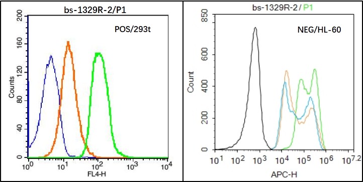
ZO-1/TJP1 Polyclonal AntibodyBS-1329R
ApplicationsFlow Cytometry, ImmunoFluorescence, Western Blot, ELISA, ImmunoCytoChemistry, ImmunoHistoChemistry, ImmunoHistoChemistry Frozen, ImmunoHistoChemistry Paraffin
ReactivityBovine, Canine, Chicken, Guinea Pig, Goat, Human, Mouse, Porcine, Rat, Sheep
TargetTJP1
- SizePrice