


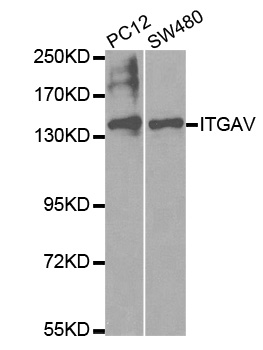
Figure 1. Western blot analysis of Integrin alpha V/ITGAV using anti-Integrin alpha V/ITGAV antibody (A01561-2). Electrophoresis was performed on a 5-20% SDS-PAGE gel at 70V (Stacking gel) / 90V (Resolving gel) for 2-3 hours. The sample well of each lane was loaded with 50ug of sample under reducing conditions. Lane 1: human A549 whole cell lysates, Lane 2: human A431 whole cell lysates. After Electrophoresis, proteins were transferred to a Nitrocellulose membrane at 150mA for 50-90 minutes. Blocked the membrane with 5% Non-fat Milk/ TBS for 1.5 hour at RT. The membrane was incubated with rabbit anti-Integrin alpha V/ITGAV antigen affinity purified polyclonal antibody (Catalog # A01561-2) at 0.25 microg/mL overnight at 4°C, then washed with TBS-0.1%Tween 3 times with 5 minutes each and probed with a goat anti-rabbit IgG-HRP secondary antibody at a dilution of 1:10000 for 1.5 hour at RT. The signal is developed using an Enhanced Chemiluminescent detection (ECL) kit (Catalog # EK1002) with Tanon 5200 system. A specific band was detected for Integrin alpha V/ITGAV at approximately 130-140KD. The expected band size for Integrin alpha V/ITGAV is at 130KD.
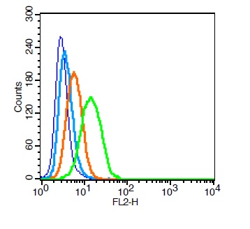
. Overlay histogram showing Raji cells stained with A01561-2 (Blue line). The cells were fixed with 4% paraformaldehyde and blocked with 10% normal goat serum. And then incubated with rabbit anti-Integrin alpha V/ITGAV Antibody (A01561-2,1microg/1x106 cells) for 30 min at 20°C. DyLight®488 conjugated goat anti-rabbit IgG (BA1127, 5-10microg/1x106 cells) was used as secondary antibody for 30 minutes at 20°C. Isotype control antibody (Green line) was rabbit IgG (1microg/1x106) used under the same conditions. Unlabelled sample without incubation with primary antibody and secondary antibody (Red line) was used as a blank control.")
Figure 1. Western blot analysis of Integrin alpha V/ITGAV using anti-Integrin alpha V/ITGAV antibody (A01561-2). Electrophoresis was performed on a 5-20% SDS-PAGE gel at 70V (Stacking gel) / 90V (Resolving gel) for 2-3 hours. The sample well of each lane was loaded with 50ug of sample under reducing conditions. Lane 1: human A549 whole cell lysates, Lane 2: human A431 whole cell lysates. After Electrophoresis, proteins were transferred to a Nitrocellulose membrane at 150mA for 50-90 minutes. Blocked the membrane with 5% Non-fat Milk/ TBS for 1.5 hour at RT. The membrane was incubated with rabbit anti-Integrin alpha V/ITGAV antigen affinity purified polyclonal antibody (Catalog # A01561-2) at 0.25 microg/mL overnight at 4°C, then washed with TBS-0.1%Tween 3 times with 5 minutes each and probed with a goat anti-rabbit IgG-HRP secondary antibody at a dilution of 1:10000 for 1.5 hour at RT. The signal is developed using an Enhanced Chemiluminescent detection (ECL) kit (Catalog # EK1002) with Tanon 5200 system. A specific band was detected for Integrin alpha V/ITGAV at approximately 130-140KD. The expected band size for Integrin alpha V/ITGAV is at 130KD.
Anti-Integrin alpha V/ITGAV Antibody Picoband(r)
A01561-2-CARRIER-FREE
ApplicationsFlow Cytometry, Western Blot, ELISA
Product group Antibodies
ReactivityHuman
TargetITGAV
Overview
- SupplierBoster Bio
- Product NameAnti-Integrin alpha V/ITGAV Antibody Picoband(r)
- Delivery Days Customer9
- ApplicationsFlow Cytometry, Western Blot, ELISA
- CertificationResearch Use Only
- ClonalityPolyclonal
- Concentration500 ug/ml
- Gene ID3685
- Target nameITGAV
- Target descriptionintegrin subunit alpha V
- Target synonymsCD51, MSK8, VNRA, VTNR, integrin alpha-V, antigen identified by monoclonal antibody L230, integrin alphaVbeta3, integrin, alpha V (vitronectin receptor, alpha polypeptide, antigen CD51), vitronectin receptor subunit alpha
- HostRabbit
- IsotypeIgG
- Protein IDP06756
- Protein NameIntegrin alpha-V
- Scientific DescriptionBoster Bio Anti-Integrin alpha V/ITGAV Antibody Picoband® catalog # A01561-2. Tested in ELISA, Flow Cytometry, WB applications. This antibody reacts with Human. The brand Picoband indicates this is a premium antibody that guarantees superior quality, high affinity, and strong signals with minimal background in Western blot applications. Only our best-performing antibodies are designated as Picoband, ensuring unmatched performance.
- ReactivityHuman
- Storage Instruction-20°C,2°C to 8°C
- UNSPSC12352203
Related products
Product group Antibodies
Anti-ITGAV AntibodyA37579
ApplicationsWestern Blot, ImmunoHistoChemistry
ReactivityHuman, Mouse, Rat
- SizePrice
Product group Antibodies
Anti-ITGAV Antibody144-02091
ApplicationsWestern Blot, ImmunoHistoChemistry
ReactivityHuman, Mouse, Rat
TargetITGAV
- SizePrice
Product group Antibodies

ReactivityHuman
TargetITGAV
- SizePrice
Product group Antibodies

Anti-alpha-V integrin [EM01309]Ab00887-1.1
ApplicationsFlow Cytometry, Western Blot, ELISA, ImmunoHistoChemistry
ReactivityHuman
TargetITGAV
- SizePrice
Product group Antibodies
References
ApplicationsFlow Cytometry, ImmunoFluorescence, Western Blot, ELISA, ImmunoCytoChemistry, ImmunoHistoChemistry, ImmunoHistoChemistry Frozen, ImmunoHistoChemistry Paraffin
ReactivityHuman, Mouse, Rat
TargetITGAV
- SizePrice
Product group Antibodies
ITGAV AntibodyCSB-PA003047
ApplicationsWestern Blot, ELISA, ImmunoHistoChemistry
ReactivityHuman, Mouse
TargetITGAV
- SizePrice
Product group Antibodies
Itgav Polyclonal AntibodyCAC11195
ApplicationsImmunoFluorescence, Western Blot, ELISA, ImmunoHistoChemistry
TargetITGAV
- SizePrice
Product group Antibodies

ApplicationsFlow Cytometry, ImmunoPrecipitation, ELISA, ImmunoHistoChemistry, ImmunoHistoChemistry Frozen
ReactivityHuman
TargetITGAV
- SizePrice
Product group Antibodies
ApplicationsWestern Blot, ELISA, ImmunoHistoChemistry, ImmunoHistoChemistry Paraffin
ReactivityHuman, Mouse
TargetITGAV
- SizePrice

Product group Antibodies
Anti-ITGAV AntibodyHPA004856
ApplicationsImmunoCytoChemistry, ImmunoHistoChemistry
ReactivityHuman
TargetITGAV
- SizePrice