

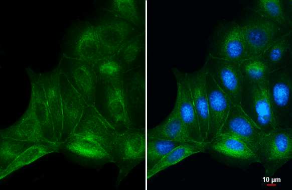
beta Catenin antibody detects beta Catenin protein at cell membrane and cytoplasm by immunofluorescent analysis. Sample: MDCK cells were fixed in 4% paraformaldehyde at RT for 15 min. Green: beta Catenin stained by beta Catenin antibody (GTX632676) diluted at 1:1000.
 diluted at 1:200. Antigen Retrieval: Citrate buffer, pH 6.0, 15 min")
 diluted at 1:200. Antigen Retrieval: Citrate buffer, pH 6.0, 15 min")
 was separated by 7.5% SDS-PAGE, and the membrane was blotted with beta Catenin antibody (GTX632676) diluted at 1:1000. The HRP-conjugated anti-mouse IgG antibody (GTX213111-01) was used to detect the primary antibody.")
 was separated by 7.5% SDS-PAGE, and the membrane was blotted with beta Catenin antibody (GTX632676) diluted at 1:1000. The HRP-conjugated anti-mouse IgG antibody (GTX213111-01) was used to detect the primary antibody, and the signal was developed with Trident ECL plus-Enhanced.")
![Wild-type (WT) and beta Catenin knockout (KO) 293T cell extracts (30 μg) were separated by 7.5% SDS-PAGE, and the membrane was blotted with beta Catenin antibody [GT3171] (GTX632676) diluted at 1:500. The HRP-conjugated anti-mouse IgG antibody (GTX213111-01) was used to detect the primary antibody, and the signal was developed with Trident ECL plus-Enhanced.](https://www.genetex.com/upload/website/prouct_img/normal/GTX632676/GTX632676_42289_20200110_WB_KO_watermark_w_23061202_546.webp "Wild-type (WT) and beta Catenin knockout (KO) 293T cell extracts (30 μg) were separated by 7.5% SDS-PAGE, and the membrane was blotted with beta Catenin antibody [GT3171] (GTX632676) diluted at 1:500. The HRP-conjugated anti-mouse IgG antibody (GTX213111-01) was used to detect the primary antibody, and the signal was developed with Trident ECL plus-Enhanced.")
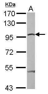
![Various whole cell extracts (30 μg) were separated by 7.5% SDS-PAGE, and the membrane was blotted with beta Catenin antibody [GT3171] (GTX632676) diluted at 1:1000.](https://www.genetex.com/upload/website/prouct_img/normal/GTX632676/GTX632676_42289_20151119_WB_w_23061202_742.webp "Various whole cell extracts (30 μg) were separated by 7.5% SDS-PAGE, and the membrane was blotted with beta Catenin antibody [GT3171] (GTX632676) diluted at 1:1000.")
 were separated by 7.5% SDS-PAGE, and the membranes were blotted with beta Catenin antibody (GTX632676) diluted at 1:500 and competitor's antibody (# Highly Cited Antibody ) diluted at 1:500. The HRP-conjugated anti-mouse IgG antibody (GTX213111-01) was used to detect the primary antibody. *The competitor is not affiliated with GeneTex and does not endorse this product.")
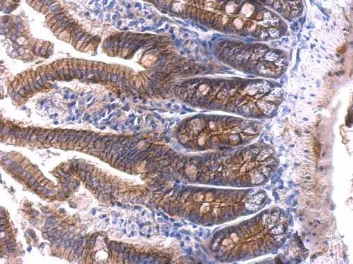
![beta Catenin antibody [GT3171] detects beta Catenin protein at cell membrane and cytoplasm in mouse intestine by immunohistochemical analysis. Sample: Paraffin-embedded mouse intestine. beta Catenin antibody [GT3171] (GTX632676) diluted at 1:400.
Antigen Retrieval: Citrate buffer, pH 6.0, 15 min](https://www.genetex.com/upload/website/prouct_img/normal/GTX632676/GTX632676_42289_20160616_IHC-P_M_w_23061202_328.webp "beta Catenin antibody [GT3171] detects beta Catenin protein at cell membrane and cytoplasm in mouse intestine by immunohistochemical analysis. Sample: Paraffin-embedded mouse intestine. beta Catenin antibody [GT3171] (GTX632676) diluted at 1:400.
Antigen Retrieval: Citrate buffer, pH 6.0, 15 min")
![Non-transfected (–) and transfected (+) HeLa whole cell extracts (30 μg) were separated by 7.5% SDS-PAGE, and the membrane was blotted with beta Catenin antibody [GT3171] (GTX632676) diluted at 1:1000.](https://www.genetex.com/upload/website/prouct_img/normal/GTX632676/GTX632676_42289_20160623_WB_shRNA_watermark_w_23061202_834.webp "Non-transfected (–) and transfected (+) HeLa whole cell extracts (30 μg) were separated by 7.5% SDS-PAGE, and the membrane was blotted with beta Catenin antibody [GT3171] (GTX632676) diluted at 1:1000.")
beta Catenin antibody detects beta Catenin protein at cell membrane and cytoplasm by immunofluorescent analysis. Sample: MDCK cells were fixed in 4% paraformaldehyde at RT for 15 min. Green: beta Catenin stained by beta Catenin antibody (GTX632676) diluted at 1:1000.
beta Catenin antibody [GT3171]
GTX632676
ApplicationsImmunoFluorescence, Western Blot, ImmunoCytoChemistry, ImmunoHistoChemistry, ImmunoHistoChemistry Paraffin
Product group Antibodies
ReactivityCanine, Feline, Human, Mouse
TargetCTNNB1
Overview
- SupplierGeneTex
- Product Namebeta Catenin antibody [GT3171]
- Delivery Days Customer9
- Application Supplier NoteWB: 1:500-1:3000. IHC-P: 1:100-1:1000. *Optimal dilutions/concentrations should be determined by the researcher.Not tested in other applications.
- ApplicationsImmunoFluorescence, Western Blot, ImmunoCytoChemistry, ImmunoHistoChemistry, ImmunoHistoChemistry Paraffin
- CertificationResearch Use Only
- ClonalityMonoclonal
- Clone IDGT3171
- Concentration1.18 mg/ml
- ConjugateUnconjugated
- Gene ID1499
- Target nameCTNNB1
- Target descriptioncatenin beta 1
- Target synonymsCTNNB, EVR7, MRD19, NEDSDV, armadillo, catenin beta-1, catenin (cadherin-associated protein), beta 1, 88kDa
- HostMouse
- IsotypeIgG1
- Protein IDP35222
- Protein NameCatenin beta-1
- Scientific DescriptionBeta-catenin is an adherens junction protein. Adherens junctions (AJs; also called the zonula adherens) are critical for the establishment and maintenance of epithelial layers, such as those lining organ surfaces. AJs mediate adhesion between cells, communicate a signal that neighboring cells are present, and anchor the actin cytoskeleton. In serving these roles, AJs regulate normal cell growth and behavior. At several stages of embryogenesis, wound healing, and tumor cell metastasis, cells form and leave epithelia. This process, which involves the disruption and reestablishment of epithelial cell-cell contacts, may be regulated by the disassembly and assembly of AJs. AJs may also function in the transmission of the contact inhibition signal, which instructs cells to stop dividing once an epithelial sheet is complete.[supplied by OMIM]
- ReactivityCanine, Feline, Human, Mouse
- Storage Instruction-20°C or -80°C,2°C to 8°C
- UNSPSC12352203
References
- Hsu CH, Liu IF, Kuo HF, et al. miR-29a-3p/THBS2 Axis Regulates PAH-Induced Cardiac Fibrosis. Int J Mol Sci. 2021,22(19). doi: 10.3390/ijms221910574Read this paper
Datasheet
Related products
Product group Antibodies

Anti-beta catenin [15B8]Ab01655-1.1
ApplicationsFlow Cytometry, ImmunoFluorescence, Western Blot, ImmunoHistoChemistry, Other Application
ReactivityBovine, Canine, Chicken, Human, Mouse, Rat
TargetCTNNB1
- SizePrice
Product group Antibodies
Anti-CTNNB1 Antibody144-11512
ApplicationsImmunoPrecipitation, Western Blot, ImmunoHistoChemistry
ReactivityHuman, Mouse, Rat
TargetCTNNB1
- SizePrice

Product group Antibodies
Anti-beta Catenin/CTNNB1 Antibody Picoband(r)A00004-CARRIER-FREE
ApplicationsFlow Cytometry, ImmunoFluorescence, Western Blot, ELISA, ImmunoCytoChemistry, ImmunoHistoChemistry
ReactivityHuman, Mouse, Rat
TargetCTNNB1
- SizePrice
Product group Antibodies
References
ApplicationsWestern Blot
ReactivityHuman, Mouse, Rat
TargetCTNNB1
- SizePrice
![WB analysis of HeLa whole cell lysate using GTX00948 beta Catenin antibody [GT1186]. Dilution : 1:1000](https://www.genetex.com/upload/website/prouct_img/normal/GTX00948/GTX00948_20200327_WB_1_w_23053121_214.webp)
Product group Antibodies
beta Catenin antibody [GT1186]GTX00948
ApplicationsImmunoFluorescence, Western Blot, ImmunoCytoChemistry, ImmunoHistoChemistry, ImmunoHistoChemistry Paraffin
ReactivityHuman, Mouse, Rat
TargetCTNNB1
- SizePrice
Product group Antibodies
References
beta Catenin antibodyGTX101254
ApplicationsImmunoFluorescence, Western Blot, ImmunoCytoChemistry, ImmunoHistoChemistry, ImmunoHistoChemistry Paraffin
ReactivityHuman, Mouse
TargetCTNNB1
- SizePrice
![Immunohistochemical analysis of paraffin-embedded zebrafish tissue, using beta Catenin antibody [N1N2-2], N-term (GTX101435) at 1:300 dilution.](https://www.genetex.com/upload/website/prouct_img/normal/GTX101435/GTX101435_39876_IHC_22111422_262.webp)
Product group Antibodies
References
ApplicationsFlow Cytometry, ImmunoFluorescence, ImmunoPrecipitation, Western Blot, ChIP Chromatin ImmunoPrecipitation, ImmunoCytoChemistry, ImmunoHistoChemistry, ImmunoHistoChemistry Frozen, ImmunoHistoChemistry Paraffin
ReactivityCanine, Feline, Human, Mouse, Rabbit, Rat, Zebra Fish
TargetCTNNB1
- SizePrice
Product group Antibodies
beta Catenin antibody, InternalGTX10342
ApplicationsWestern Blot, ChIP Chromatin ImmunoPrecipitation
ReactivityHuman
TargetCTNNB1
- SizePrice
Product group Antibodies
beta Catenin antibodyGTX110594
ApplicationsWestern Blot
ReactivityHuman
TargetCTNNB1
- SizePrice
Product group Antibodies
References
ApplicationsWestern Blot, ELISA
ReactivityChicken, Human, Mouse, Rat
TargetCTNNB1
- SizePrice