

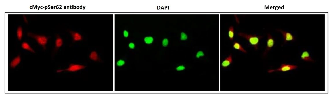
ICC/IF analysis of HeLa cells using GTX00684 c-Myc (phospho Ser62) antibody [33A12E10]. Dilution : 1:2000 in 1% BSA in PBS at 4 degree C, overnight Fixation : 4% PFA, overnight Permeabilization : 0.25% Triton X-100 in PBS for 10 min
![ICC/IF analysis of HeLa cells using GTX00684 c-Myc (phospho Ser62) antibody [33A12E10] (Green, Left) or proximity Ligation Analysis with anti-c-Myc pS62 and CIP2A antibodies, association of c-Myc pS62 with CIP2A (Right, red) in nuclei (DAPI, blue).](https://www.genetex.com/upload/website/prouct_img/normal/GTX00684/GTX00684_20191104_ICC-IF_1_w_23053121_552.webp "ICC/IF analysis of HeLa cells using GTX00684 c-Myc (phospho Ser62) antibody [33A12E10] (Green, Left) or proximity Ligation Analysis with anti-c-Myc pS62 and CIP2A antibodies, association of c-Myc pS62 with CIP2A (Right, red) in nuclei (DAPI, blue).")
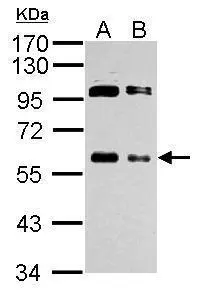
![WB analysis of various samples using GTX00684 c-Myc (phospho Ser62) antibody [33A12E10]. Scrambled siRNA : AGS cells transfected with scrambled siRNA Negative control siRNA : AGS cells transfected with negative control siRNA
Myc1 : AGS cells transfected with c-Myc siRNA](https://www.genetex.com/upload/website/prouct_img/normal/GTX00684/GTX00684_20191104_WB_w_23053121_749.webp "WB analysis of various samples using GTX00684 c-Myc (phospho Ser62) antibody [33A12E10]. Scrambled siRNA : AGS cells transfected with scrambled siRNA Negative control siRNA : AGS cells transfected with negative control siRNA
Myc1 : AGS cells transfected with c-Myc siRNA")
ICC/IF analysis of HeLa cells using GTX00684 c-Myc (phospho Ser62) antibody [33A12E10]. Dilution : 1:2000 in 1% BSA in PBS at 4 degree C, overnight Fixation : 4% PFA, overnight Permeabilization : 0.25% Triton X-100 in PBS for 10 min
c-Myc (phospho Ser62) antibody [33A12E10]
GTX00684
ApplicationsFlow Cytometry, ImmunoFluorescence, ImmunoPrecipitation, Western Blot, ELISA, ImmunoCytoChemistry, ImmunoHistoChemistry, ImmunoHistoChemistry Paraffin
Product group Antibodies
ReactivityHuman, Mouse
TargetMYC
Overview
- SupplierGeneTex
- Product Namec-Myc (phospho Ser62) antibody [33A12E10]
- Delivery Days Customer9
- Application Supplier NoteWB: ~1 microg/ml. ICC/IF: 0.5-1 microg/ml. IHC-P: 5 microg/ml. FACS: using 1 microg for 106 cells.. *Optimal dilutions/concentrations should be determined by the researcher.Not tested in other applications.
- ApplicationsFlow Cytometry, ImmunoFluorescence, ImmunoPrecipitation, Western Blot, ELISA, ImmunoCytoChemistry, ImmunoHistoChemistry, ImmunoHistoChemistry Paraffin
- CertificationResearch Use Only
- ClonalityMonoclonal
- Clone ID33A12E10
- Concentration1 mg/ml
- ConjugateUnconjugated
- Gene ID4609
- Target nameMYC
- Target descriptionMYC proto-oncogene, bHLH transcription factor
- Target synonymsMRTL, MYCC, bHLHe39, c-Myc, myc proto-oncogene protein, avian myelocytomatosis viral oncogene homolog, class E basic helix-loop-helix protein 39, myc-related translation/localization regulatory factor, proto-oncogene c-Myc, transcription factor p64, v-myc avian myelocytomatosis viral oncogene homolog, v-myc myelocytomatosis viral oncogene homolog
- HostMouse
- IsotypeIgG2b
- Protein IDP01106
- Protein NameMyc proto-oncogene protein
- Scientific DescriptionThis gene is a proto-oncogene and encodes a nuclear phosphoprotein that plays a role in cell cycle progression, apoptosis and cellular transformation. The encoded protein forms a heterodimer with the related transcription factor MAX. This complex binds to the E box DNA consensus sequence and regulates the transcription of specific target genes. Amplification of this gene is frequently observed in numerous human cancers. Translocations involving this gene are associated with Burkitt lymphoma and multiple myeloma in human patients. There is evidence to show that translation initiates both from an upstream, in-frame non-AUG (CUG) and a downstream AUG start site, resulting in the production of two isoforms with distinct N-termini. [provided by RefSeq, Aug 2017]
- ReactivityHuman, Mouse
- Storage Instruction-20°C or -80°C,2°C to 8°C
- UNSPSC12352203
References
- Lee CC, Ho KH, Huang TW, et al. A regulatory loop among CD276, miR-29c-3p, and Myc exists in cancer cells against natural killer cell cytotoxicity. Life Sci. 2021,277:119438. doi: 10.1016/j.lfs.2021.119438Read this paper
- Myant K, Qiao X, Halonen T, et al. Serine 62-Phosphorylated MYC Associates with Nuclear Lamins and Its Regulation by CIP2A Is Essential for Regenerative Proliferation. Cell Rep. 2015,12(6):1019-31. doi: 10.1016/j.celrep.2015.07.003Read this paper
- Tibbitts DC, Escamilla-Powers JR, Zhang X, et al. Studying c-Myc serine 62 phosphorylation in leukemia cells: concern over antibody cross-reactivity. Blood. 2012,119(22):5334-5. doi: 10.1182/blood-2012-03-414532Read this paper
- Mathiasen DP, Egebjerg C, Andersen SH, et al. Identification of a c-Jun N-terminal kinase-2-dependent signal amplification cascade that regulates c-Myc levels in ras transformation. Oncogene. 2012,31(3):390-401. doi: 10.1038/onc.2011.230Read this paper
- Tallents RH, Jarvis R. Provisional restorations. Part Two. Oral Health. 1988,78(2):29-33.Read this paper
Datasheet
Related products
Product group Antibodies

Anti-myc [8]Ab02771-10.0
ApplicationsImmunoPrecipitation, Western Blot, ImmunoHistoChemistry
ReactivityHuman, Mouse, Rat
TargetMYC
- SizePrice
Product group Antibodies
Anti-MYC Antibody144-01309
ApplicationsWestern Blot
ReactivityHuman, Mouse, Rat
TargetMYC
- SizePrice

Product group Antibodies
Anti-c-Myc/MYC Antibody Picoband(r)A00026-1-CARRIER-FREE
ApplicationsFlow Cytometry, Western Blot, ELISA
ReactivityHuman, Mouse, Rat
TargetMYC
- SizePrice
Product group Antibodies
c-Myc antibody (HRP)GTX12213
ApplicationsImmunoFluorescence, Western Blot, ELISA, ImmunoCytoChemistry, ImmunoHistoChemistry
ReactivityHuman
TargetMYC
- SizePrice
![Untreated (–) and treated (+) HepG2 whole cell extracts (30 μg) were separated by 10% SDS-PAGE, and the membrane was blotted with c-Myc (phospho Thr58/Ser62) antibody [SZ02-06] (GTX01137) diluted at 1:500. The HRP-conjugated anti-rabbit IgG antibody (GTX213110-01) was used to detect the primary antibody.](https://www.genetex.com/upload/website/prouct_img/normal/GTX01137/GTX01137_HK1219_20200228_WB_treatment_MG132_w_23053121_696.webp)
Product group Antibodies
ApplicationsWestern Blot
ReactivityHuman
TargetMYC
- SizePrice
![WB analysis of mouse ovary tissue lysate using GTX02877 c-Myc antibody [GT1265]. Dilution : 1:1000 Loading : 25μg per lane](https://www.genetex.com/upload/website/prouct_img/normal/GTX02877/GTX02877_20210226_WB_2_w_23053123_529.webp)
Product group Antibodies
c-Myc antibody [GT1265]GTX02877
ApplicationsImmunoFluorescence, Western Blot, ImmunoCytoChemistry
ReactivityHuman, Mouse
TargetMYC
- SizePrice
![Untreated (–) and treated (+) NIH-3T3 whole cell extracts (30 μg) were separated by 10% SDS-PAGE, and the membrane was blotted with c-Myc (phospho Ser62) antibody [GT1295] (GTX03207) diluted at 1:500. The HRP-conjugated anti-rabbit IgG antibody (GTX213110-01) was used to detect the primary antibody.](https://www.genetex.com/upload/website/prouct_img/normal/GTX03207/GTX03207_4000001533_20210716_WB_M_CIP_w_23053123_302.webp)
Product group Antibodies
ApplicationsWestern Blot
ReactivityHuman, Mouse
TargetMYC
- SizePrice
![IHC-P analysis of human colon adenocarcinoma tissue using GTX04712 c-Myc antibody [9E10]. Antigen retrieval : Citrate Buffer pH 6.0_x000D_](https://www.genetex.com/upload/website/prouct_img/normal/GTX04712/GTX04712_20240325_IHC-P_24032422_493.webp)
Product group Antibodies
c-Myc antibody [9E10]GTX04712
ApplicationsImmunoHistoChemistry, ImmunoHistoChemistry Paraffin
ReactivityHuman
TargetMYC
- SizePrice
Product group Antibodies
References
c-Myc antibodyGTX103436
ApplicationsImmunoFluorescence, ImmunoPrecipitation, Western Blot, ChIP Chromatin ImmunoPrecipitation, ImmunoCytoChemistry, ImmunoHistoChemistry, ImmunoHistoChemistry Frozen, ImmunoHistoChemistry Paraffin
ReactivityHuman, Mouse, Xenopus
TargetMYC
- SizePrice
![FACS (intracellular staining) analysis of transfected LST-1-c-Myc in HEK-293 cells compared with nontransfected HEK-293 cells using GTX10826 c-Myc antibody [9E10] (FITC).](https://www.genetex.com/upload/website/prouct_img/normal/GTX10826/GTX10826_20191028_FACS_1_w_23060120_474.webp)
Product group Antibodies
References
c-Myc antibody [9E10] (FITC)GTX10826
ApplicationsFlow Cytometry
ReactivityHuman
TargetMYC
- SizePrice