

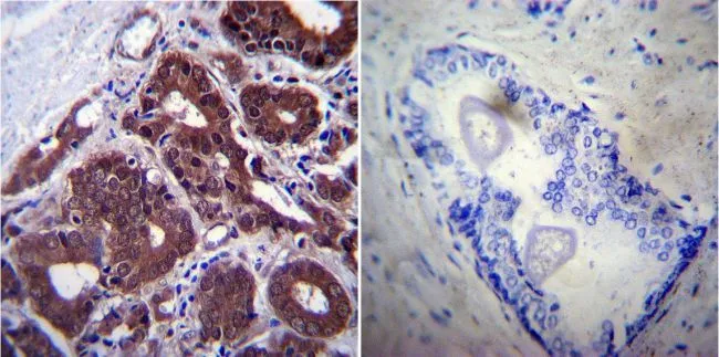
IHC-P analysis of human prostate carcinoma tissue using GTX25442 Hsp70 antibody [2A4]. Left : Primary antibody Right : Negative control without primary antibody Antigen retrieval : heat induced antigen retrieval was performed using 10mM sodium citrate (pH6.0) buffer, microwaved for 8-15 minutes Dilution : 1:200
![ICC/IF analysis of NIH-3T3 cells using GTX25442 Hsp70 antibody [2A4]. Cells were probed without (right) or with(left) an antibody. Green : Primary antibody Blue : Nuclei Red : Actin Fixation : formaldehyde Dilution : 1:100 overnight at 4 oC](https://www.genetex.com/upload/website/prouct_img/normal/GTX25442/GTX25442_649_ICC-IF_w_23060722_636.webp "ICC/IF analysis of NIH-3T3 cells using GTX25442 Hsp70 antibody [2A4]. Cells were probed without (right) or with(left) an antibody. Green : Primary antibody Blue : Nuclei Red : Actin Fixation : formaldehyde Dilution : 1:100 overnight at 4 oC")
![ICC/IF analysis of NIH-3T3 cells using GTX25442 Hsp70 antibody [2A4]. Cells were probed without (left) or with(right) an antibody. Green : Primary antibody Blue : Nuclei Red : Actin Fixation : Formalin Permeabilization : 0.1% Triton X-100 in TBS for 5-10 minute Dilution : 1:200 and incubated overnight in a humidified chamber](https://www.genetex.com/upload/website/prouct_img/normal/GTX25442/GTX25442_648_ICC-IF_w_23060722_208.webp "ICC/IF analysis of NIH-3T3 cells using GTX25442 Hsp70 antibody [2A4]. Cells were probed without (left) or with(right) an antibody. Green : Primary antibody Blue : Nuclei Red : Actin Fixation : Formalin Permeabilization : 0.1% Triton X-100 in TBS for 5-10 minute Dilution : 1:200 and incubated overnight in a humidified chamber")
![IP analysis of HeLa cell lysates using GTX25442 Hsp70 antibody [2A4]. IP reaction : 2μg antibody / 500μg lysate Dilution : 1:1000](https://www.genetex.com/upload/website/prouct_img/normal/GTX25442/GTX25442_1443_IP_w_23060722_983.webp "IP analysis of HeLa cell lysates using GTX25442 Hsp70 antibody [2A4]. IP reaction : 2μg antibody / 500μg lysate Dilution : 1:1000")
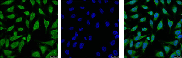
![ICC/IF analysis of HeLa and NIH3T3 cells using GTX25442 Hsp70 antibody [2A4]. Green : Primary antibody Blue : Nuclei Fixation : Formalin Permeabilization : 0.1% Triton X-100 in TBS for 10 minutes Dilution : 1:50 for at least 1 hour at room temperature](https://www.genetex.com/upload/website/prouct_img/normal/GTX25442/GTX25442_652_ICC-IF_w_23060722_850.webp "ICC/IF analysis of HeLa and NIH3T3 cells using GTX25442 Hsp70 antibody [2A4]. Green : Primary antibody Blue : Nuclei Fixation : Formalin Permeabilization : 0.1% Triton X-100 in TBS for 10 minutes Dilution : 1:50 for at least 1 hour at room temperature")
![IHC-P analysis of human breast tissue using GTX25442 Hsp70 antibody [2A4]. Left : Primary antibody Right : Negative control without primary antibody Antigen retrieval : heat induced antigen retrieval was performed using 10mM sodium citrate (pH6.0) buffer, microwaved for 8-15 minutes Dilution : 1:200](https://www.genetex.com/upload/website/prouct_img/normal/GTX25442/GTX25442_1287_IHC-P_w_23060722_416.webp "IHC-P analysis of human breast tissue using GTX25442 Hsp70 antibody [2A4]. Left : Primary antibody Right : Negative control without primary antibody Antigen retrieval : heat induced antigen retrieval was performed using 10mM sodium citrate (pH6.0) buffer, microwaved for 8-15 minutes Dilution : 1:200")
![ICC/IF analysis of HeLa cells using GTX25442 Hsp70 antibody [2A4]. Cells were probed without (left) or with(right) an antibody. Green : Primary antibody Blue : Nuclei Red : Actin Fixation : Formalin Permeabilization : 0.1% Triton X-100 in TBS for 5-10 minute Dilution : 1:100 incubated overnight in a humidified chamber](https://www.genetex.com/upload/website/prouct_img/normal/GTX25442/GTX25442_651_ICC-IF_w_23060722_822.webp "ICC/IF analysis of HeLa cells using GTX25442 Hsp70 antibody [2A4]. Cells were probed without (left) or with(right) an antibody. Green : Primary antibody Blue : Nuclei Red : Actin Fixation : Formalin Permeabilization : 0.1% Triton X-100 in TBS for 5-10 minute Dilution : 1:100 incubated overnight in a humidified chamber")
![WB analysis of 50 ug of the indicated whole cell lysates using GTX25442 Hsp70 antibody [2A4]. Dilution : 1:1000](https://www.genetex.com/upload/website/prouct_img/normal/GTX25442/GTX25442_1776_WB_w_23060722_241.webp "WB analysis of 50 ug of the indicated whole cell lysates using GTX25442 Hsp70 antibody [2A4]. Dilution : 1:1000")
![WB analysis of 30 ug of HEK293T whole cell lysate per well using GTX25442 Hsp70 antibody [2A4]. Dilution : 1:1000](https://www.genetex.com/upload/website/prouct_img/normal/GTX25442/GTX25442_1775_WB_w_23060722_478.webp "WB analysis of 30 ug of HEK293T whole cell lysate per well using GTX25442 Hsp70 antibody [2A4]. Dilution : 1:1000")
![IHC-P analysis of human tonsil tissue using GTX25442 Hsp70 antibody [2A4]. Left : Primary antibody Right : Negative control without primary antibody Antigen retrieval : heat induced antigen retrieval was performed using 10mM sodium citrate (pH6.0) buffer, microwaved for 8-15 minutes Dilution : 1:200](https://www.genetex.com/upload/website/prouct_img/normal/GTX25442/GTX25442_1288_IHC-P_w_23060722_497.webp "IHC-P analysis of human tonsil tissue using GTX25442 Hsp70 antibody [2A4]. Left : Primary antibody Right : Negative control without primary antibody Antigen retrieval : heat induced antigen retrieval was performed using 10mM sodium citrate (pH6.0) buffer, microwaved for 8-15 minutes Dilution : 1:200")
IHC-P analysis of human prostate carcinoma tissue using GTX25442 Hsp70 antibody [2A4]. Left : Primary antibody Right : Negative control without primary antibody Antigen retrieval : heat induced antigen retrieval was performed using 10mM sodium citrate (pH6.0) buffer, microwaved for 8-15 minutes Dilution : 1:200
Hsp70 antibody [2A4]
GTX25442
ApplicationsImmunoFluorescence, ImmunoPrecipitation, Western Blot, ImmunoCytoChemistry, ImmunoHistoChemistry, ImmunoHistoChemistry Paraffin, Neutralisation/Blocking
Product group Antibodies
ReactivityAmphibian, Avian, Drosophila, Fish, Human, Mouse, Primate, Rat, Yeast, Zebra Fish
TargetHSPA1A
Overview
- SupplierGeneTex
- Product NameHsp70 antibody [2A4]
- Delivery Days Customer9
- Application Supplier NoteWB: 1:1000 - 1:2500. ICC/IF: 1:100-1:200. IHC-P: 1:200. IP: 2 microg. *Optimal dilutions/concentrations should be determined by the researcher.Not tested in other applications.
- ApplicationsImmunoFluorescence, ImmunoPrecipitation, Western Blot, ImmunoCytoChemistry, ImmunoHistoChemistry, ImmunoHistoChemistry Paraffin, Neutralisation/Blocking
- CertificationResearch Use Only
- ClonalityMonoclonal
- Clone ID2A4
- Concentration3.5 mg/ml
- ConjugateUnconjugated
- Gene ID3303
- Target nameHSPA1A
- Target descriptionheat shock protein family A (Hsp70) member 1A
- Target synonymsHEL-S-103, HSP70, HSP70-1, HSP70-1A, HSP70-2, HSP70.1, HSP70.2, HSP70I, HSP72, HSPA1, heat shock 70 kDa protein 1A, HSP70-1/HSP70-2, HSP70.1/HSP70.2, Heat shock 70 kDa protein 1B, Heat shock 70 kDa protein 2, dnaK-type molecular chaperone HSP70-1, epididymis secretory protein Li 103, epididymis secretory sperm binding protein, heat shock 70 kDa protein 1, heat shock 70 kDa protein 1/2, heat shock 70 kDa protein 1A/1B, heat shock 70kD protein 1A, heat shock 70kDa protein 1A, heat shock protein family A member 1A, heat shock-induced protein
- HostMouse
- IsotypeIgM
- Protein IDP0DMV8
- Protein NameHeat shock 70 kDa protein 1A
- ReactivityAmphibian, Avian, Drosophila, Fish, Human, Mouse, Primate, Rat, Yeast, Zebra Fish
- Storage Instruction-20°C or -80°C,2°C to 8°C
- UNSPSC41116161
References
- Critical Effects on Akt Signaling in Adult Zebrafish Brain Following Alterations in Light Exposure. Moore NS et al., 2021 Mar 12, CellsRead this paper
Datasheet
Related products
Product group Antibodies
Anti-HSP70 AntibodyA81956
ApplicationsWestern Blot, ELISA, ImmunoHistoChemistry
- SizePrice
Product group Antibodies

Anti-HSP70 [cmhsp70.1 (C92F3B1)]Ab03197-1.1
ApplicationsFlow Cytometry, ImmunoFluorescence, Western Blot, ELISA, ImmunoHistoChemistry
ReactivityHuman
TargetHSPA1A
- SizePrice

Product group Antibodies
ApplicationsImmunoFluorescence, Western Blot, ELISA, ImmunoHistoChemistry
ReactivityHuman, Mouse, Rat
TargetHSPA1A
- SizePrice
Product group Antibodies

HSPA1A AntibodyLS-C763520
ApplicationsWestern Blot, ImmunoHistoChemistry
ReactivityHuman
TargetHSPA1A
- SizePrice

Product group Antibodies
Anti-HSPA1A AntibodyHPA052504
ApplicationsImmunoCytoChemistry
ReactivityHuman
TargetHSPA1A
- SizePrice
Product group Antibodies
HSPA1L Monoclonal AntibodyCSB-MA000197
ApplicationsImmunoFluorescence, Western Blot, ELISA, ImmunoHistoChemistry
ReactivityHuman, Mouse, Rat
TargetHSPA1A
- SizePrice
![WB analysis of 20ul of gill tissue lysates from the salt marsh mussel, Guekensia demissa isolated from various coves in Rhode Island (indicated above the lanes) using GTX25439 Hsp70 antibody [3A3]. Dilution : 1:1000](https://www.genetex.com/upload/website/prouct_img/normal/GTX25439/GTX25439_1773_WB_w_23060722_300.webp)
Product group Antibodies
References
Hsp70 antibody [3A3]GTX25439
ApplicationsGel Shift Assay, Flow Cytometry, ImmunoFluorescence, ImmunoPrecipitation, Western Blot, ELISA, ImmunoCytoChemistry, ImmunoHistoChemistry, ImmunoHistoChemistry Paraffin, Neutralisation/Blocking
ReactivityAmphibian, Avian, Drosophila, Fish, Human, Insect, Monkey, Molluscs, Mouse, Plant, Porcine, Primate, Rat, Yeast, Other Species
TargetHSPA1A
- SizePrice
![IHC-P analysis of human breast tissue using GTX25444 Hsp70 antibody [4G4]. Left : Primary antibody Right : Negative control without primary antibody Antigen retrieval : heat induced antigen retrieval was performed using 10mM sodium citrate (pH6.0) buffer, microwaved for 8-15 minutes Dilution : 1:50](https://www.genetex.com/upload/website/prouct_img/normal/GTX25444/GTX25444_1290_IHC-P_w_23060722_915.webp)
Product group Antibodies
Hsp70 antibody [4G4]GTX25444
ApplicationsFlow Cytometry, ImmunoFluorescence, ImmunoPrecipitation, Western Blot, ImmunoCytoChemistry, ImmunoHistoChemistry, ImmunoHistoChemistry Paraffin, Neutralisation/Blocking, Other Application
ReactivityHuman, Mouse, Primate
TargetHSPA1A
- SizePrice
Product group Antibodies
HSPA1A Polyclonal AntibodyCAC15126
ApplicationsImmunoFluorescence, Western Blot, ELISA, ImmunoHistoChemistry
TargetHSPA1A
- SizePrice