

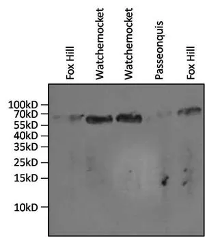
WB analysis of 20ul of gill tissue lysates from the salt marsh mussel, Guekensia demissa isolated from various coves in Rhode Island (indicated above the lanes) using GTX25439 Hsp70 antibody [3A3]. Dilution : 1:1000
![IP analysis of HeLa cell lysates using GTX25439 Hsp70 antibody [3A3]. IP reaction : 2μg antibody / 500μg lysate Dilution : 1:1000](https://www.genetex.com/upload/website/prouct_img/normal/GTX25439/GTX25439_1442_IP_w_23060722_270.webp "IP analysis of HeLa cell lysates using GTX25439 Hsp70 antibody [3A3]. IP reaction : 2μg antibody / 500μg lysate Dilution : 1:1000")
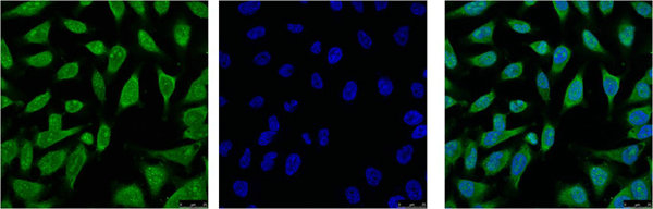
![ICC/IF analysis of HeLa and NIH3T3 cells using GTX25439 Hsp70 antibody [3A3]. Green : Primary antibody Blue : Nuclei Fixation : Formalin Permeabilization : 0.1% Triton X-100 in TBS for 10 minutes Dilution : 1:50 for at least 1 hour at room temperature](https://www.genetex.com/upload/website/prouct_img/normal/GTX25439/GTX25439_643_ICC-IF_w_23060722_144.webp "ICC/IF analysis of HeLa and NIH3T3 cells using GTX25439 Hsp70 antibody [3A3]. Green : Primary antibody Blue : Nuclei Fixation : Formalin Permeabilization : 0.1% Triton X-100 in TBS for 10 minutes Dilution : 1:50 for at least 1 hour at room temperature")
![ICC/IF analysis of NIH-3T3 cells using GTX25439 Hsp70 antibody [3A3]. Cells were probed without (right) or with(left) an antibody. Green : Primary antibody Blue : Nuclei Red : Actin Fixation : formaldehyde Dilution : 1:200 overnight at 4oC](https://www.genetex.com/upload/website/prouct_img/normal/GTX25439/GTX25439_642_ICC-IF_w_23060722_978.webp "ICC/IF analysis of NIH-3T3 cells using GTX25439 Hsp70 antibody [3A3]. Cells were probed without (right) or with(left) an antibody. Green : Primary antibody Blue : Nuclei Red : Actin Fixation : formaldehyde Dilution : 1:200 overnight at 4oC")
![ICC/IF analysis of MCF-7 cells using GTX25439 Hsp70 antibody [3A3]. Cells were probed without (right) or with(left) an antibody. Green : Primary antibody Blue : Nuclei Red : Actin Fixation : formaldehyde Dilution : 1:200 overnight at 4oC](https://www.genetex.com/upload/website/prouct_img/normal/GTX25439/GTX25439_641_ICC-IF_w_23060722_342.webp "ICC/IF analysis of MCF-7 cells using GTX25439 Hsp70 antibody [3A3]. Cells were probed without (right) or with(left) an antibody. Green : Primary antibody Blue : Nuclei Red : Actin Fixation : formaldehyde Dilution : 1:200 overnight at 4oC")
![ICC/IF analysis of HeLa cells using GTX25439 Hsp70 antibody [3A3]. Cells were probed without (right) or with(left) an antibody. Green : Primary antibody Blue : Nuclei Red : Actin Fixation : formaldehyde Dilution : 1:200 overnight at 4oC](https://www.genetex.com/upload/website/prouct_img/normal/GTX25439/GTX25439_644_ICC-IF_w_23060722_953.webp "ICC/IF analysis of HeLa cells using GTX25439 Hsp70 antibody [3A3]. Cells were probed without (right) or with(left) an antibody. Green : Primary antibody Blue : Nuclei Red : Actin Fixation : formaldehyde Dilution : 1:200 overnight at 4oC")
![IHC-P analysis of human breast carcinoma tissue using GTX25439 Hsp70 antibody [3A3]. Left : Primary antibody Right : Negative control without primary antibody Antigen retrieval : heat induced antigen retrieval was performed using 10mM sodium citrate (pH6.0) buffer, microwaved for 8-15 minutes Dilution : 1:50](https://www.genetex.com/upload/website/prouct_img/normal/GTX25439/GTX25439_1283_IHC-P_w_23060722_400.webp "IHC-P analysis of human breast carcinoma tissue using GTX25439 Hsp70 antibody [3A3]. Left : Primary antibody Right : Negative control without primary antibody Antigen retrieval : heat induced antigen retrieval was performed using 10mM sodium citrate (pH6.0) buffer, microwaved for 8-15 minutes Dilution : 1:50")
![IHC-P analysis of human testis tissue using GTX25439 Hsp70 antibody [3A3]. Left : Primary antibody Right : Negative control without primary antibody Antigen retrieval : heat induced antigen retrieval was performed using 10mM sodium citrate (pH6.0) buffer, microwaved for 8-15 minutes Dilution : 1:200](https://www.genetex.com/upload/website/prouct_img/normal/GTX25439/GTX25439_1284_IHC-P_w_23060722_525.webp "IHC-P analysis of human testis tissue using GTX25439 Hsp70 antibody [3A3]. Left : Primary antibody Right : Negative control without primary antibody Antigen retrieval : heat induced antigen retrieval was performed using 10mM sodium citrate (pH6.0) buffer, microwaved for 8-15 minutes Dilution : 1:200")
![IHC-P analysis of human tonsil tissue using GTX25439 Hsp70 antibody [3A3]. Left : Primary antibody Right : Negative control without primary antibody Antigen retrieval : heat induced antigen retrieval was performed using 10mM sodium citrate (pH6.0) buffer, microwaved for 8-15 minutes Dilution : 1:20](https://www.genetex.com/upload/website/prouct_img/normal/GTX25439/GTX25439_1285_IHC-P_w_23060722_675.webp "IHC-P analysis of human tonsil tissue using GTX25439 Hsp70 antibody [3A3]. Left : Primary antibody Right : Negative control without primary antibody Antigen retrieval : heat induced antigen retrieval was performed using 10mM sodium citrate (pH6.0) buffer, microwaved for 8-15 minutes Dilution : 1:20")
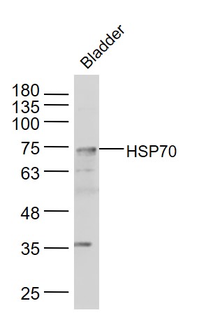
![WB analysis of 50 ug of the indicated whole cell lysates using GTX25439 Hsp70 antibody [3A3]. Dilution : 1:1000](https://www.genetex.com/upload/website/prouct_img/normal/GTX25439/GTX25439_1772_WB_w_23060722_977.webp "WB analysis of 50 ug of the indicated whole cell lysates using GTX25439 Hsp70 antibody [3A3]. Dilution : 1:1000")
WB analysis of 20ul of gill tissue lysates from the salt marsh mussel, Guekensia demissa isolated from various coves in Rhode Island (indicated above the lanes) using GTX25439 Hsp70 antibody [3A3]. Dilution : 1:1000
Hsp70 antibody [3A3]
GTX25439
ApplicationsGel Shift Assay, Flow Cytometry, ImmunoFluorescence, ImmunoPrecipitation, Western Blot, ELISA, ImmunoCytoChemistry, ImmunoHistoChemistry, ImmunoHistoChemistry Paraffin, Neutralisation/Blocking
Product group Antibodies
ReactivityAmphibian, Avian, Drosophila, Fish, Human, Insect, Monkey, Molluscs, Mouse, Plant, Porcine, Primate, Rat, Yeast, Other Species
TargetHSPA1A
Overview
- SupplierGeneTex
- Product NameHsp70 antibody [3A3]
- Delivery Days Customer9
- Application Supplier NoteWB: 1:1000 - 1:5000. ICC/IF: 1:100. IHC-P: 1:200. IP: 2 microl. *Optimal dilutions/concentrations should be determined by the researcher.Not tested in other applications.
- ApplicationsGel Shift Assay, Flow Cytometry, ImmunoFluorescence, ImmunoPrecipitation, Western Blot, ELISA, ImmunoCytoChemistry, ImmunoHistoChemistry, ImmunoHistoChemistry Paraffin, Neutralisation/Blocking
- CertificationResearch Use Only
- ClonalityMonoclonal
- Clone ID3A3
- Concentration1 mg/ml
- ConjugateUnconjugated
- Gene ID3303
- Target nameHSPA1A
- Target descriptionheat shock protein family A (Hsp70) member 1A
- Target synonymsHEL-S-103, HSP70, HSP70-1, HSP70-1A, HSP70-2, HSP70.1, HSP70.2, HSP70I, HSP72, HSPA1, heat shock 70 kDa protein 1A, HSP70-1/HSP70-2, HSP70.1/HSP70.2, Heat shock 70 kDa protein 1B, Heat shock 70 kDa protein 2, dnaK-type molecular chaperone HSP70-1, epididymis secretory protein Li 103, epididymis secretory sperm binding protein, heat shock 70 kDa protein 1, heat shock 70 kDa protein 1/2, heat shock 70 kDa protein 1A/1B, heat shock 70kD protein 1A, heat shock 70kDa protein 1A, heat shock protein family A member 1A, heat shock-induced protein
- HostMouse
- IsotypeIgG1
- Protein IDP0DMV8
- Protein NameHeat shock 70 kDa protein 1A
- ReactivityAmphibian, Avian, Drosophila, Fish, Human, Insect, Monkey, Molluscs, Mouse, Plant, Porcine, Primate, Rat, Yeast, Other Species
- Storage Instruction-20°C or -80°C,2°C to 8°C
- UNSPSC41116161
References
- Identification of the gene encoding the TATA box-binding protein-associated factor 1 (TAF1) and its putative role in the heat shock response in the protozoan parasite Entamoeba histolytica. Avendano-Borromeo B et al., 2019 Feb, Parasitol ResRead this paper
- Caveolin-1 Secreted from Adipose Tissues and Adipocytes Functions as an Adipogenesis Enhancer. Chang CC et al., 2017 Nov, Obesity (Silver Spring)Read this paper
- Extracellular Hsp70 Enhances Mesoangioblast Migration via an Autocrine Signaling Pathway. Barreca MM et al., 2017 Jul, J Cell PhysiolRead this paper
- HSP70 regulates the function of mitotic centrosomes. Fang CT et al., 2016 Oct, Cell Mol Life SciRead this paper
- Immunohistochemical distribution of heat shock protein 70 and proliferating cell nuclear antigen in mouse placenta at different gestational stages. Ozaydin T et al., 2016 Apr, Microsc Res TechRead this paper
- A calreticulin-dependent nuclear export signal is involved in the regulation of liver receptor homologue-1 protein folding. Yang FM et al., 2015 Oct 15, Biochem JRead this paper
- Hyperbaric oxygen preconditioning induces tolerance against oxidative injury and oxygen-glucose deprivation by up-regulating heat shock protein 32 in rat spinal neurons. Huang G et al., 2014, PLoS OneRead this paper
Datasheet
Related products
Product group Antibodies
Anti-HSP70 AntibodyA81956
ApplicationsWestern Blot, ELISA, ImmunoHistoChemistry
- SizePrice
Product group Antibodies

Anti-HSP70 [cmhsp70.1 (C92F3B1)]Ab03197-1.1
ApplicationsFlow Cytometry, ImmunoFluorescence, Western Blot, ELISA, ImmunoHistoChemistry
ReactivityHuman
TargetHSPA1A
- SizePrice
Product group Antibodies
Anti-HSP70 Antibody130-10052
ApplicationsELISA
ReactivityHuman
TargetHSPA1A
- SizePrice

Product group Antibodies
References
HSP70 Polyclonal AntibodyBS-0126R
ApplicationsFlow Cytometry, ImmunoFluorescence, Western Blot, ELISA, ImmunoCytoChemistry, ImmunoHistoChemistry, ImmunoHistoChemistry Frozen, ImmunoHistoChemistry Paraffin
ReactivityBovine, Chicken, Human, Mouse, Rabbit, Rat, Sheep
TargetHSPA1A
- SizePrice
Product group Antibodies
HSPA1L Monoclonal AntibodyCSB-MA000197
ApplicationsImmunoFluorescence, Western Blot, ELISA, ImmunoHistoChemistry
ReactivityHuman, Mouse, Rat
TargetHSPA1A
- SizePrice
Product group Antibodies
HSPA1A Polyclonal AntibodyCAC15126
ApplicationsImmunoFluorescence, Western Blot, ELISA, ImmunoHistoChemistry
TargetHSPA1A
- SizePrice
![IHC-P analysis of human tonsil tissue using GTX22787 Hsp70 antibody [5A5]. Left : Primary antibody Right : Negative control without primary antibody Antigen retrieval : heat induced antigen retrieval was performed using 10mM sodium citrate (pH6.0) buffer, microwaved for 8-15 minutes Dilution : 1:200](https://www.genetex.com/upload/website/prouct_img/normal/GTX22787/GTX22787_1112_IHC-P_w_23060620_584.webp)
Product group Antibodies
References
Hsp70 antibody [5A5]GTX22787
ApplicationsGel Shift Assay, ImmunoFluorescence, ImmunoPrecipitation, Western Blot, ELISA, ImmunoCytoChemistry, ImmunoHistoChemistry, ImmunoHistoChemistry Frozen, ImmunoHistoChemistry Paraffin, Neutralisation/Blocking
ReactivityAmphibian, Avian, Bovine, Drosophila, Fish, Human, Molluscs, Mouse, Rabbit, Rat, Yeast, Other Species
TargetHSPA1A
- SizePrice
Product group Antibodies
References
Hsp70 antibody [W27]GTX23148
ApplicationsFlow Cytometry, ImmunoFluorescence, ImmunoPrecipitation, Western Blot, ImmunoCytoChemistry, ImmunoHistoChemistry, ImmunoHistoChemistry Paraffin
ReactivityBovine, Human, Monkey
TargetHSPA1A
- SizePrice