

ICC/IF analysis of HeLa cells using GTX15779 PLK1 antibody [13E8]. Fixation : 4% paraformaldehyde Permeabilization : 0.1% Triton X-100 for 10 minute Dilution : 3 μg/ml in 0.1% BSA and incubated for overnight at 4oC
![ICC/IF analysis of HeLa cells using GTX15779 PLK1 antibody [13E8]. Cells were probed without (right) or with(left) an antibody. Green : Primary antibody Blue : Nuclei Red : Actin Fixation : formaldehyde Dilution : 1:20 overnight at 4oC](https://www.genetex.com/upload/website/prouct_img/normal/GTX15779/GTX15779_308_ICC-IF_w_23060620_598.webp "ICC/IF analysis of HeLa cells using GTX15779 PLK1 antibody [13E8]. Cells were probed without (right) or with(left) an antibody. Green : Primary antibody Blue : Nuclei Red : Actin Fixation : formaldehyde Dilution : 1:20 overnight at 4oC")
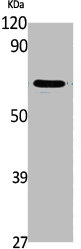
![WB analysis of PLK1 knockdown HeLa cells (lane 3), scrambled siRNA transfected cells (lane 2) and untransfected cells (lane 1) using GTX15779 PLK1 antibody [13E8]. Dilution : 1 μg/ml](https://www.genetex.com/upload/website/prouct_img/normal/GTX15779/GTX15779_1531_WB_w_23060620_113.webp "WB analysis of PLK1 knockdown HeLa cells (lane 3), scrambled siRNA transfected cells (lane 2) and untransfected cells (lane 1) using GTX15779 PLK1 antibody [13E8]. Dilution : 1 μg/ml")
![WB analysis of whole cell extracts (30μg) of HeLa (Lane 1), treated with Nocodazole (500nM for 18 hrs) (Lane2), SW-480 (Lane 3), treated with Nocodazole (500nM for 18 hrs) (Lane 4), COLO 205 (Lane 5), HCT 116 (Lane 6), K-562 (Lane 7), MDA-MB-231 (Lane 8), HepG2 (Lane 9), A549 (Lane 10) using GTX15779 PLK1 antibody [13E8]. Dilution : 1:1000](https://www.genetex.com/upload/website/prouct_img/normal/GTX15779/GTX15779_1533_WB_w_23060620_644.webp "WB analysis of whole cell extracts (30μg) of HeLa (Lane 1), treated with Nocodazole (500nM for 18 hrs) (Lane2), SW-480 (Lane 3), treated with Nocodazole (500nM for 18 hrs) (Lane 4), COLO 205 (Lane 5), HCT 116 (Lane 6), K-562 (Lane 7), MDA-MB-231 (Lane 8), HepG2 (Lane 9), A549 (Lane 10) using GTX15779 PLK1 antibody [13E8]. Dilution : 1:1000")
![ICC/IF analysis of U251 cells using GTX15779 PLK1 antibody [13E8]. Cells were probed without (right) or with(left) an antibody. Green : Primary antibody Blue : Nuclei Red : Actin Fixation : formaldehyde Dilution : 1:20 overnight at 4oC](https://www.genetex.com/upload/website/prouct_img/normal/GTX15779/GTX15779_310_ICC-IF_w_23060620_511.webp "ICC/IF analysis of U251 cells using GTX15779 PLK1 antibody [13E8]. Cells were probed without (right) or with(left) an antibody. Green : Primary antibody Blue : Nuclei Red : Actin Fixation : formaldehyde Dilution : 1:20 overnight at 4oC")
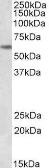
![WB analysis of untreated or Nocodazole (100nM, 48 hours) treated U2OS lysate from non-targeting control or PLK1 siRNA transfected U2OS cells using GTX15779 PLK1 antibody [13E8]. Dilution : 1:1000 Loading : 25 μg](https://www.genetex.com/upload/website/prouct_img/normal/GTX15779/GTX15779_1532_WB_w_23060620_993.webp "WB analysis of untreated or Nocodazole (100nM, 48 hours) treated U2OS lysate from non-targeting control or PLK1 siRNA transfected U2OS cells using GTX15779 PLK1 antibody [13E8]. Dilution : 1:1000 Loading : 25 μg")
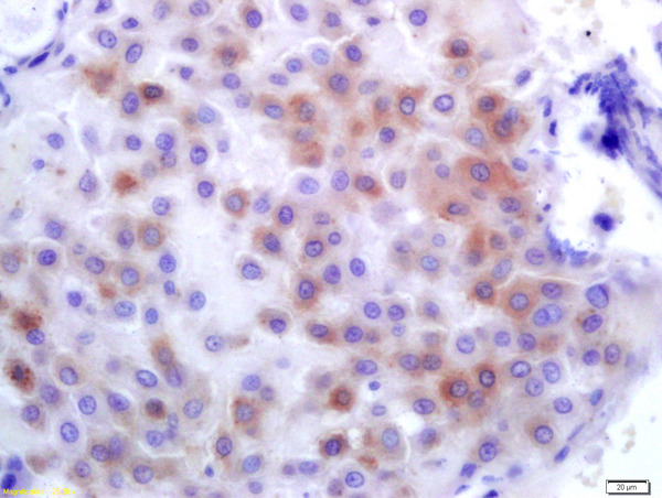
![IHC-P analysis of human colon tissue using GTX15779 PLK1 antibody [13E8]. Left : Primary antibody Right : Negative control without primary antibody Antigen retrieval : heat induced antigen retrieval was performed using 10mM sodium citrate (pH6.0) buffer, microwaved for 8-15 minutes Dilution : 1:200](https://www.genetex.com/upload/website/prouct_img/normal/GTX15779/GTX15779_1039_IHC-P_w_23060620_913.webp "IHC-P analysis of human colon tissue using GTX15779 PLK1 antibody [13E8]. Left : Primary antibody Right : Negative control without primary antibody Antigen retrieval : heat induced antigen retrieval was performed using 10mM sodium citrate (pH6.0) buffer, microwaved for 8-15 minutes Dilution : 1:200")
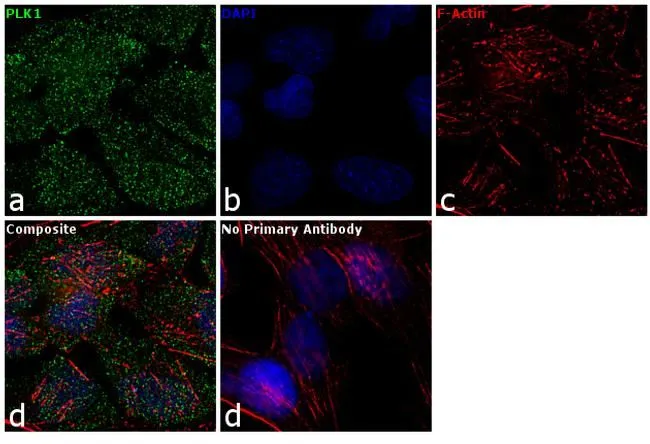
![ICC/IF analysis of HeLa cells using GTX15779 PLK1 antibody [13E8]. Progression of cells through subsequent phases of the cell cycle results in differential localization of PLK1 in spindle poles, kinetochores and cleavage furrow. Green : Primary antibody Blue : Nuclei Red : Actin Fixation : 4% paraformaldehyde Permeabilization : 0.1% Triton X-100 for 10 minute Dilution : 3 μg/ml in 0.1% BSA and incubated for overnight at 4oC](https://www.genetex.com/upload/website/prouct_img/normal/GTX15779/GTX15779_307_ICC-IF_w_23060620_237.webp "ICC/IF analysis of HeLa cells using GTX15779 PLK1 antibody [13E8]. Progression of cells through subsequent phases of the cell cycle results in differential localization of PLK1 in spindle poles, kinetochores and cleavage furrow. Green : Primary antibody Blue : Nuclei Red : Actin Fixation : 4% paraformaldehyde Permeabilization : 0.1% Triton X-100 for 10 minute Dilution : 3 μg/ml in 0.1% BSA and incubated for overnight at 4oC")
![ICC/IF analysis of WiDr cells using GTX15779 PLK1 antibody [13E8]. Cells were probed without (right) or with(left) an antibody. Green : Primary antibody Blue : Nuclei Red : Actin Fixation : formaldehyde Dilution : 1:20 overnight at 4oC](https://www.genetex.com/upload/website/prouct_img/normal/GTX15779/GTX15779_309_ICC-IF_w_23060620_406.webp "ICC/IF analysis of WiDr cells using GTX15779 PLK1 antibody [13E8]. Cells were probed without (right) or with(left) an antibody. Green : Primary antibody Blue : Nuclei Red : Actin Fixation : formaldehyde Dilution : 1:20 overnight at 4oC")
ICC/IF analysis of HeLa cells using GTX15779 PLK1 antibody [13E8]. Fixation : 4% paraformaldehyde Permeabilization : 0.1% Triton X-100 for 10 minute Dilution : 3 μg/ml in 0.1% BSA and incubated for overnight at 4oC
PLK1 antibody [13E8]
GTX15779
ApplicationsImmunoFluorescence, ImmunoPrecipitation, Western Blot, ELISA, ImmunoCytoChemistry, ImmunoHistoChemistry, ImmunoHistoChemistry Paraffin
Product group Antibodies
ReactivityHuman, Mouse, Rat
TargetPLK1
Overview
- SupplierGeneTex
- Product NamePLK1 antibody [13E8]
- Delivery Days Customer9
- Application Supplier NoteWB: 1:1,000. ICC/IF: 3 microg/ml. IHC-P: 1:20. *Optimal dilutions/concentrations should be determined by the researcher.Not tested in other applications.
- ApplicationsImmunoFluorescence, ImmunoPrecipitation, Western Blot, ELISA, ImmunoCytoChemistry, ImmunoHistoChemistry, ImmunoHistoChemistry Paraffin
- CertificationResearch Use Only
- ClonalityMonoclonal
- Concentration1 mg/ml
- ConjugateUnconjugated
- Gene ID5347
- Target namePLK1
- Target descriptionpolo like kinase 1
- Target synonymsPLK, STPK13, serine/threonine-protein kinase PLK1, PLK-1, cell cycle regulated protein kinase, polo (Drosophia)-like kinase, serine/threonine-protein kinase 13
- HostMouse
- IsotypeIgG2b
- Protein IDP53350
- Protein NameSerine/threonine-protein kinase PLK1
- Scientific DescriptionThe Ser/Thr protein kinase encoded by this gene belongs to the CDC5/Polo subfamily. It is highly expressed during mitosis and elevated levels are found in many different types of cancer. Depletion of this protein in cancer cells dramatically inhibited cell proliferation and induced apoptosis; hence, it is a target for cancer therapy. [provided by RefSeq, Sep 2015]
- ReactivityHuman, Mouse, Rat
- Storage Instruction-20°C or -80°C,2°C to 8°C
- UNSPSC41116161
Datasheet
Related products
Product group Antibodies
Anti-PLK1 AntibodyA85042
ApplicationsWestern Blot, ELISA
ReactivityHuman
- SizePrice

Product group Antibodies
Anti-PLK1 AntibodyAMAB91515
ApplicationsWestern Blot
ReactivityHuman
TargetPLK1
- SizePrice
Product group Antibodies
References
PLK1 Polyclonal AntibodyBS-3535R
ApplicationsImmunoFluorescence, Western Blot, ELISA, ImmunoCytoChemistry, ImmunoHistoChemistry, ImmunoHistoChemistry Frozen, ImmunoHistoChemistry Paraffin
ReactivityCanine, Human, Mouse, Porcine, Rabbit, Rat
TargetPLK1
- SizePrice
Product group Antibodies

Goat anti-PLK1EB06969
ApplicationsWestern Blot, ELISA
ReactivityCanine, Human, Mouse, Rat
TargetPLK1
- SizePrice
Product group Antibodies
PLK1 AntibodyCSB-PA004668
ApplicationsWestern Blot, ELISA
ReactivityHuman, Mouse, Rat
TargetPLK1
- SizePrice
![IHC-P analysis of human colon tissue using GTX14210 PLK1 antibody [Mixed clones]. Right : Primary antibody Left : Negative control without primary antibody Antigen retireval : 10mM sodium citrate (pH 6.0), microwaved for 8-15 min Dilution : 1:20](https://www.genetex.com/upload/website/prouct_img/normal/GTX14210/GTX14210_20191203_IHC-P_20_w_23060620_745.webp)
Product group Antibodies
PLK1 antibody [Mixed clones]GTX14210
ApplicationsFlow Cytometry, ImmunoFluorescence, ImmunoPrecipitation, Western Blot, ELISA, ImmunoCytoChemistry, ImmunoHistoChemistry, ImmunoHistoChemistry Paraffin
ReactivityHuman, Monkey, Mouse, Rat, Xenopus
TargetPLK1
- SizePrice
Product group Antibodies
PLK1 (phospho Tyr217) antibodyGTX18156
ApplicationsWestern Blot
ReactivityHuman
TargetPLK1
- SizePrice
![IHC-P analysis of squamous cell carcinoma of oropharangeal tissue using GTX01927 PLK1 antibody [MJS1]. Note the intense nuclear and cytoplasmic staining of a proportion of proliferating malignant cells.](https://www.genetex.com/upload/website/prouct_img/normal/GTX01927/GTX01927_20200811_IHC-P_73_w_23053121_314.webp)
Product group Antibodies
PLK1 antibody [MJS1]GTX01927
ApplicationsImmunoHistoChemistry, ImmunoHistoChemistry Paraffin
ReactivityHuman
TargetPLK1
- SizePrice
Product group Antibodies
PLK1 (phospho Thr210) antibodyGTX03368
ApplicationsImmunoFluorescence, Western Blot, ELISA, ImmunoCytoChemistry, ImmunoHistoChemistry, ImmunoHistoChemistry Paraffin
ReactivityHuman, Mouse
TargetPLK1
- SizePrice
Product group Antibodies

PLK1 / PLK-1 Antibody (Internal)LS-C368472
ApplicationsImmunoFluorescence, Western Blot, ImmunoCytoChemistry, ImmunoHistoChemistry, ImmunoHistoChemistry Paraffin
ReactivityBovine, Human, Monkey, Mouse, Porcine, Zebra Fish
TargetPLK1
- SizePrice