

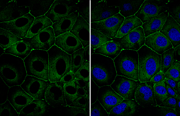
ZO-1 antibody detects ZO-1 protein at cell membrane and cytoplasm by immunofluorescent analysis. Sample: MDCK cells were fixed in 4% paraformaldehyde at RT for 15 min. Green: ZO-1 stained by ZO-1 antibody (GTX108592) diluted at 1:1000.
 diluted at 1:1000.")
 was separated by 5% SDS-PAGE, and the membrane was blotted with ZO-1 antibody (GTX108592) diluted at 1:1000. The HRP-conjugated anti-rabbit IgG antibody (GTX213110-01) was used to detect the primary antibody.")
 diluted at 1:500. Antigen Retrieval: Citrate buffer, pH 6.0, 15 min")
 and transfected (+) HeLa whole cell extracts (30 μg) were separated by 5% SDS-PAGE, and the membrane was blotted with ZO-1 antibody (GTX108592) diluted at 1:500. The HRP-conjugated anti-rabbit IgG antibody (GTX213110-01) was used to detect the primary antibody.")
 diluted at 1:500.
Antigen Retrieval: Citrate buffer, pH 6.0, 15 min")
 were separated by 5% SDS-PAGE, and the membrane was blotted with ZO-1 antibody (GTX108592) diluted at 1:1000.")
 was separated by 5% SDS-PAGE, and the membrane was blotted with ZO-1 antibody (GTX108592) diluted at 1:500. The HRP-conjugated anti-rabbit IgG antibody (GTX213110-01) was used to detect the primary antibody.")


ZO-1 antibody detects ZO-1 protein at cell membrane and cytoplasm by immunofluorescent analysis. Sample: MDCK cells were fixed in 4% paraformaldehyde at RT for 15 min. Green: ZO-1 stained by ZO-1 antibody (GTX108592) diluted at 1:1000.
ZO-1 antibody
GTX108592
ApplicationsImmunoFluorescence, Western Blot, ImmunoCytoChemistry, ImmunoHistoChemistry, ImmunoHistoChemistry Frozen, ImmunoHistoChemistry Paraffin
Product group Antibodies
ReactivityBovine, Canine, Human, Monkey, Mouse, Porcine
TargetTJP1
Overview
- SupplierGeneTex
- Product NameZO-1 antibody
- Delivery Days Customer9
- Application Supplier NoteWB: 1:500-1:3000. IHC-P: 1:100-1:1000. *Optimal dilutions/concentrations should be determined by the researcher.Not tested in other applications.
- ApplicationsImmunoFluorescence, Western Blot, ImmunoCytoChemistry, ImmunoHistoChemistry, ImmunoHistoChemistry Frozen, ImmunoHistoChemistry Paraffin
- CertificationResearch Use Only
- ClonalityPolyclonal
- Concentration0.19 mg/ml
- ConjugateUnconjugated
- Gene ID7082
- Target nameTJP1
- Target descriptiontight junction protein 1
- Target synonymsZO-1, tight junction protein 1, tight junction protein ZO-1, zona occludens 1, zonula occludens 1 protein
- HostRabbit
- IsotypeIgG
- Protein IDQ07157
- Protein NameTight junction protein 1
- Scientific DescriptionThis gene encodes a protein located on a cytoplasmic membrane surface of intercellular tight junctions. The encoded protein may be involved in signal transduction at cell-cell junctions. Two transcript variants encoding distinct isoforms have been identified for this gene. [provided by RefSeq]
- ReactivityBovine, Canine, Human, Monkey, Mouse, Porcine
- Storage Instruction-20°C or -80°C,2°C to 8°C
- UNSPSC12352203
References
- Han P, Lei Y, Liu J, et al. Cell adhesion molecule BVES functions as a suppressor of tumor cells extrusion in hepatocellular carcinoma metastasis. Cell Commun Signal. 2022,20(1):149. doi: 10.1186/s12964-022-00962-9Read this paper
- Wei MF, Cheng CH, Wen SY, et al. Atorvastatin Attenuates Radiotherapy-Induced Intestinal Damage through Activation of Autophagy and Antioxidant Effects. Oxid Med Cell Longev. 2022,2022:7957255. doi: 10.1155/2022/7957255Read this paper
- Fang PH, Lai YY, Chen CL, et al. Cobalt protoporphyrin promotes human keratinocyte migration under hyperglycemic conditions. Mol Med. 2022,28(1):71. doi: 10.1186/s10020-022-00499-0Read this paper
- De Tomi E, Campagnari R, Orlandi E, et al. Upregulation of miR-34a-5p, miR-20a-3p and miR-29a-3p by Onconase in A375 Melanoma Cells Correlates with the Downregulation of Specific Onco-Proteins. Int J Mol Sci. 2022,23(3). doi: 10.3390/ijms23031647Read this paper
- Barbian ME, Owens JA, Naudin CR, et al. Butyrate supplementation to pregnant mice elicits cytoprotection against colonic injury in the offspring. Pediatr Res. 2022,92(1):125-134. doi: 10.1038/s41390-021-01767-1Read this paper
- Watanabe D, Nakagawa S, Morofuji Y, et al. Characterization of a Primate Blood-Brain Barrier Co-Culture Model Prepared from Primary Brain Endothelial Cells, Pericytes and Astrocytes. Pharmaceutics. 2021,13(9). doi: 10.3390/pharmaceutics13091484Read this paper
- Kariya Y, Oyama M, Suzuki T, et al. αvβ3 Integrin induces partial EMT independent of TGF-β signaling. Commun Biol. 2021,4(1):490. doi: 10.1038/s42003-021-02003-6Read this paper
- Fang W, Zhao P, Shen A, et al. Effects of Qing Hua Chang Yin on lipopolysaccharide‑induced intestinal epithelial tight junction injury in Caco‑2 cells. Mol Med Rep. 2021,23(3):pii: 205. doi: 10.3892/mmr.2021.11844.Read this paper
- Minamide K, Sato T, Nakanishi Y, et al. IRF2 maintains the stemness of colonic stem cells by limiting physiological stress from interferon. Sci Rep. 2020,10(1):14639. doi: 10.1038/s41598-020-71633-3Read this paper
- Bisi S, Marchesi S, Rizvi A, et al. IRSp53 controls plasma membrane shape and polarized transport at the nascent lumen in epithelial tubules. Nat Commun. 2020,11(1):3516. doi: 10.1038/s41467-020-17091-xRead this paper
Datasheet
Related products
Product group Antibodies
Anti-ZO-1 Antibody144-61634
ApplicationsImmunoFluorescence, Western Blot
ReactivityHuman, Mouse, Rat
TargetTJP1
- SizePrice
![Non-transfected (–) and transfected (+) HeLa whole cell extracts (30 μg) were separated by 5% SDS-PAGE, and the membrane was blotted with ZO-1 antibody [C3], C-term (GTX108587) diluted at 1:500.](https://www.genetex.com/upload/website/prouct_img/normal/GTX108587/GTX108587_40618_20160804_WB_shRNA_watermark_w_23060120_565.webp)
Product group Antibodies
References
ZO-1 antibody [C3], C-termGTX108587
ApplicationsWestern Blot
ReactivityHuman
TargetTJP1
- SizePrice
![ZO-1 antibody [N1N2], N-term detects ZO-1 protein at cell membrane by immunofluorescent analysis. Sample: A431 cells were fixed in ice-cold MeOH for 5 min. Green: ZO-1 stained by ZO-1 antibody [N1N2], N-term (GTX108613) diluted at 1:500.](https://www.genetex.com/upload/website/prouct_img/normal/GTX108613/GTX108613_43803_20210416_ICC_IF_w_23060120_527.webp)
Product group Antibodies
References
ZO-1 antibody [N1N2], N-termGTX108613
ApplicationsImmunoFluorescence, ImmunoPrecipitation, Western Blot, ImmunoCytoChemistry, ImmunoHistoChemistry, ImmunoHistoChemistry Frozen
ReactivityFish, Human, Mouse, Rat
TargetTJP1
- SizePrice
![Non-transfected (–) and transfected (+) HeLa whole cell extracts (30 μg) were separated by 5% SDS-PAGE, and the membrane was blotted with ZO-1 antibody [N2C1], Internal (GTX108627) diluted at 1:500. The HRP-conjugated anti-rabbit IgG antibody (GTX213110-01) was used to detect the primary antibody.](https://www.genetex.com/upload/website/prouct_img/normal/GTX108627/GTX108627_40464_20160804_WB_shRNA_watermark_w_23060120_175.webp)
Product group Antibodies
References
ZO-1 antibody [N2C1], InternalGTX108627
ApplicationsImmunoFluorescence, Western Blot, ImmunoCytoChemistry, ImmunoHistoChemistry, ImmunoHistoChemistry Frozen
ReactivityHuman, Rat
TargetTJP1
- SizePrice
![Wild-type (WT) and TJP1 knockout (KO) HeLa cell extracts (30 μg) were separated by 5% SDS-PAGE, and the membrane was blotted with ZO-1 antibody [HL1133] (GTX636399) diluted at 1:1000. The HRP-conjugated anti-rabbit IgG antibody (GTX213110-01) was used to detect the primary antibody.](https://www.genetex.com/upload/website/prouct_img/normal/GTX636399/GTX636399_44466_20220211_WB_KO_watermark_w_23061202_165.webp)
Product group Antibodies
References
ZO-1 antibody [HL1133]GTX636399
ApplicationsImmunoFluorescence, Western Blot, ImmunoCytoChemistry
ReactivityHuman
TargetTJP1
- SizePrice
![ZO-1 antibody [HL1185] detects ZO-1 protein at cell membrane by immunofluorescent analysis. Sample: A431 cells were fixed in 4% paraformaldehyde at RT for 15 min. Green: ZO-1 stained by ZO-1 antibody [HL1185] (GTX636491) diluted at 1:500. Blue: Fluoroshield with DAPI (GTX30920).](https://www.genetex.com/upload/website/prouct_img/normal/GTX636491/GTX636491_T-44445_20211126_ICC_IF_w_23061202_412.webp)
Product group Antibodies
ZO-1 antibody [HL1185]GTX636491
ApplicationsImmunoFluorescence, Western Blot, ImmunoCytoChemistry
ReactivityCanine, Feline, Human, Mouse, Rat
TargetTJP1
- SizePrice
Product group Antibodies
Tjp1 Polyclonal AntibodyCAC11695
ApplicationsImmunoFluorescence, ELISA
TargetTJP1
- SizePrice
Product group Antibodies
References
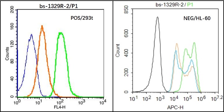
ZO-1/TJP1 Polyclonal AntibodyBS-1329R
ApplicationsFlow Cytometry, ImmunoFluorescence, Western Blot, ELISA, ImmunoCytoChemistry, ImmunoHistoChemistry, ImmunoHistoChemistry Frozen, ImmunoHistoChemistry Paraffin
ReactivityBovine, Canine, Chicken, Guinea Pig, Goat, Human, Mouse, Porcine, Rat, Sheep
TargetTJP1
- SizePrice