

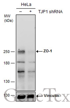
Non-transfected (–) and transfected (+) HeLa whole cell extracts (30 μg) were separated by 5% SDS-PAGE, and the membrane was blotted with ZO-1 antibody [N2C1], Internal (GTX108627) diluted at 1:500. The HRP-conjugated anti-rabbit IgG antibody (GTX213110-01) was used to detect the primary antibody.
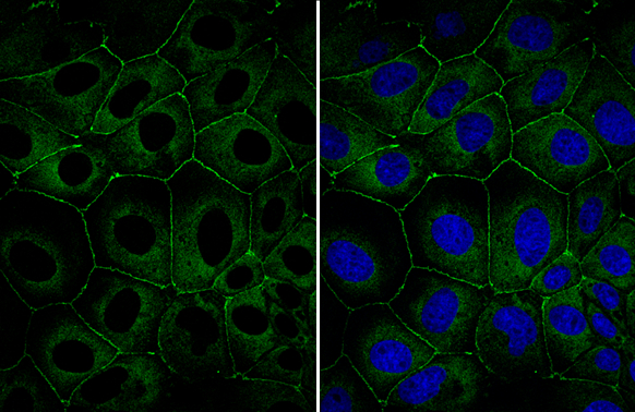
![ZO-1 antibody [N2C1], Internal detects TJP1 protein at junction by confocal immunofluorescent analysis. Sample: A431 cells were fixed in ice-cold MeOH for 5 min. Green: TJP1 protein stained by ZO-1 antibody [N2C1], Internal (GTX108627) diluted at 1:500. Blue: Hoechst 33342 staining. [Images captured by Olympus FV10i Confocal Laser Scanning Microscope]](https://www.genetex.com/upload/website/prouct_img/normal/GTX108627/GTX108627_40464_IFA_w_23060120_905.webp "ZO-1 antibody [N2C1], Internal detects TJP1 protein at junction by confocal immunofluorescent analysis. Sample: A431 cells were fixed in ice-cold MeOH for 5 min. Green: TJP1 protein stained by ZO-1 antibody [N2C1], Internal (GTX108627) diluted at 1:500. Blue: Hoechst 33342 staining. [Images captured by Olympus FV10i Confocal Laser Scanning Microscope]")
![Various whole cell extracts (30 μg) were separated by 5% SDS-PAGE, and the membrane was blotted with ZO-1 antibody [N2C1], Internal (GTX108627) diluted at 1:2000. The HRP-conjugated anti-rabbit IgG antibody (GTX213110-01) was used to detect the primary antibody.](https://www.genetex.com/upload/website/prouct_img/normal/GTX108627/GTX108627_43488_20190201_WB_w_23060120_184.webp "Various whole cell extracts (30 μg) were separated by 5% SDS-PAGE, and the membrane was blotted with ZO-1 antibody [N2C1], Internal (GTX108627) diluted at 1:2000. The HRP-conjugated anti-rabbit IgG antibody (GTX213110-01) was used to detect the primary antibody.")
![Various whole cell extracts (30 μg) were separated by 5% SDS-PAGE, and the membrane was blotted with ZO-1 antibody [N2C1], Internal (GTX108627) diluted at 1:2000. The HRP-conjugated anti-rabbit IgG antibody (GTX213110-01) was used to detect the primary antibody.](https://www.genetex.com/upload/website/prouct_img/normal/GTX108627/GTX108627_43565_20190510_WB_w_23060120_366.webp "Various whole cell extracts (30 μg) were separated by 5% SDS-PAGE, and the membrane was blotted with ZO-1 antibody [N2C1], Internal (GTX108627) diluted at 1:2000. The HRP-conjugated anti-rabbit IgG antibody (GTX213110-01) was used to detect the primary antibody.")
![Various whole cell extracts (30 μg) were separated by 5% SDS-PAGE, and the membrane was blotted with ZO-1 antibody [N2C1], Internal (GTX108627) diluted at 1:1000. The HRP-conjugated anti-rabbit IgG antibody (GTX213110-01) was used to detect the primary antibody.](https://www.genetex.com/upload/website/prouct_img/normal/GTX108627/GTX108627_45197_20231013_WB_23102401_866.webp "Various whole cell extracts (30 μg) were separated by 5% SDS-PAGE, and the membrane was blotted with ZO-1 antibody [N2C1], Internal (GTX108627) diluted at 1:1000. The HRP-conjugated anti-rabbit IgG antibody (GTX213110-01) was used to detect the primary antibody.")
.")
![Various whole cell extracts (30 μg) were separated by 5% SDS-PAGE, and the membrane was blotted with ZO-1 antibody [N2C1], Internal (GTX108627) diluted at 1:1000. The HRP-conjugated anti-rabbit IgG antibody (GTX213110-01) was used to detect the primary antibody.](https://www.genetex.com/upload/website/prouct_img/normal/GTX108627/GTX108627_45196_20240524_WB_R_24052802_531.webp "Various whole cell extracts (30 μg) were separated by 5% SDS-PAGE, and the membrane was blotted with ZO-1 antibody [N2C1], Internal (GTX108627) diluted at 1:1000. The HRP-conjugated anti-rabbit IgG antibody (GTX213110-01) was used to detect the primary antibody.")
Non-transfected (–) and transfected (+) HeLa whole cell extracts (30 μg) were separated by 5% SDS-PAGE, and the membrane was blotted with ZO-1 antibody [N2C1], Internal (GTX108627) diluted at 1:500. The HRP-conjugated anti-rabbit IgG antibody (GTX213110-01) was used to detect the primary antibody.
ZO-1 antibody [N2C1], Internal
GTX108627
ApplicationsImmunoFluorescence, Western Blot, ImmunoCytoChemistry, ImmunoHistoChemistry, ImmunoHistoChemistry Frozen
Product group Antibodies
ReactivityHuman, Rat
TargetTJP1
Overview
- SupplierGeneTex
- Product NameZO-1 antibody [N2C1], Internal
- Delivery Days Customer9
- Application Supplier NoteWB: 1:500-1:3000. ICC/IF: 1:100-1:1000. *Optimal dilutions/concentrations should be determined by the researcher.Not tested in other applications.
- ApplicationsImmunoFluorescence, Western Blot, ImmunoCytoChemistry, ImmunoHistoChemistry, ImmunoHistoChemistry Frozen
- CertificationResearch Use Only
- ClonalityPolyclonal
- Concentration0.98 mg/ml
- ConjugateUnconjugated
- Gene ID7082
- Target nameTJP1
- Target descriptiontight junction protein 1
- Target synonymsZO-1, tight junction protein 1, tight junction protein ZO-1, zona occludens 1, zonula occludens 1 protein
- HostRabbit
- IsotypeIgG
- Protein IDQ07157
- Protein NameTight junction protein 1
- Scientific DescriptionThis gene encodes a protein located on a cytoplasmic membrane surface of intercellular tight junctions. The encoded protein may be involved in signal transduction at cell-cell junctions. Two transcript variants encoding distinct isoforms have been identified for this gene. [provided by RefSeq]
- ReactivityHuman, Rat
- Storage Instruction-20°C or -80°C,2°C to 8°C
- UNSPSC12352203
References
- Wang HK, Su YT, Ho YC, et al. HDAC1 is Involved in Neuroinflammation and Blood-Brain Barrier Damage in Stroke Pathogenesis. J Inflamm Res. 2023,16:4103-4116. doi: 10.2147/JIR.S416239Read this paper
- Takino JI, Sato T, Kanetaka T, et al. RasGRP2 inhibits glyceraldehyde-derived toxic advanced glycation end-products from inducing permeability in vascular endothelial cells. Sci Rep. 2021,11(1):2959. doi: 10.1038/s41598-021-82619-0Read this paper
- Michielin F, Giobbe GG, Luni C, et al. The Microfluidic Environment Reveals a Hidden Role of Self-Organizing Extracellular Matrix in Hepatic Commitment and Organoid Formation of hiPSCs. Cell Rep. 2020,33(9):108453. doi: 10.1016/j.celrep.2020.108453Read this paper
- Hung WT, Wang CH, Lin SY, et al. Leptin protects brain from ischemia/reperfusion-induced infarction by stabilizing the blood-brain barrier to block brain infiltration by the blood-borne neutrophils. Eur J Neurosci. 2020,52(12):4890-4907. doi: 10.1111/ejn.14896Read this paper
- Huang JY, Wang YY, Lo S, et al. Visfatin Mediates Malignant Behaviors through Adipose-Derived Stem Cells Intermediary in Breast Cancer. Cancers (Basel). 2019,12(1). doi: 10.3390/cancers12010029Read this paper
Datasheet
Related products
Product group Antibodies
Anti-ZO-1 Antibody144-61634
ApplicationsImmunoFluorescence, Western Blot
ReactivityHuman, Mouse, Rat
TargetTJP1
- SizePrice
![Non-transfected (–) and transfected (+) HeLa whole cell extracts (30 μg) were separated by 5% SDS-PAGE, and the membrane was blotted with ZO-1 antibody [C3], C-term (GTX108587) diluted at 1:500.](https://www.genetex.com/upload/website/prouct_img/normal/GTX108587/GTX108587_40618_20160804_WB_shRNA_watermark_w_23060120_565.webp)
Product group Antibodies
References
ZO-1 antibody [C3], C-termGTX108587
ApplicationsWestern Blot
ReactivityHuman
TargetTJP1
- SizePrice
Product group Antibodies
References
ZO-1 antibodyGTX108592
ApplicationsImmunoFluorescence, Western Blot, ImmunoCytoChemistry, ImmunoHistoChemistry, ImmunoHistoChemistry Frozen, ImmunoHistoChemistry Paraffin
ReactivityBovine, Canine, Human, Monkey, Mouse, Porcine
TargetTJP1
- SizePrice
![ZO-1 antibody [N1N2], N-term detects ZO-1 protein at cell membrane by immunofluorescent analysis. Sample: A431 cells were fixed in ice-cold MeOH for 5 min. Green: ZO-1 stained by ZO-1 antibody [N1N2], N-term (GTX108613) diluted at 1:500.](https://www.genetex.com/upload/website/prouct_img/normal/GTX108613/GTX108613_43803_20210416_ICC_IF_w_23060120_527.webp)
Product group Antibodies
References
ZO-1 antibody [N1N2], N-termGTX108613
ApplicationsImmunoFluorescence, ImmunoPrecipitation, Western Blot, ImmunoCytoChemistry, ImmunoHistoChemistry, ImmunoHistoChemistry Frozen
ReactivityFish, Human, Mouse, Rat
TargetTJP1
- SizePrice
![Wild-type (WT) and TJP1 knockout (KO) HeLa cell extracts (30 μg) were separated by 5% SDS-PAGE, and the membrane was blotted with ZO-1 antibody [HL1133] (GTX636399) diluted at 1:1000. The HRP-conjugated anti-rabbit IgG antibody (GTX213110-01) was used to detect the primary antibody.](https://www.genetex.com/upload/website/prouct_img/normal/GTX636399/GTX636399_44466_20220211_WB_KO_watermark_w_23061202_165.webp)
Product group Antibodies
References
ZO-1 antibody [HL1133]GTX636399
ApplicationsImmunoFluorescence, Western Blot, ImmunoCytoChemistry
ReactivityHuman
TargetTJP1
- SizePrice
![ZO-1 antibody [HL1185] detects ZO-1 protein at cell membrane by immunofluorescent analysis. Sample: A431 cells were fixed in 4% paraformaldehyde at RT for 15 min. Green: ZO-1 stained by ZO-1 antibody [HL1185] (GTX636491) diluted at 1:500. Blue: Fluoroshield with DAPI (GTX30920).](https://www.genetex.com/upload/website/prouct_img/normal/GTX636491/GTX636491_T-44445_20211126_ICC_IF_w_23061202_412.webp)
Product group Antibodies
ZO-1 antibody [HL1185]GTX636491
ApplicationsImmunoFluorescence, Western Blot, ImmunoCytoChemistry
ReactivityCanine, Feline, Human, Mouse, Rat
TargetTJP1
- SizePrice
Product group Antibodies
Tjp1 Polyclonal AntibodyCAC11695
ApplicationsImmunoFluorescence, ELISA
TargetTJP1
- SizePrice
Product group Antibodies
References
ZO-1/TJP1 Polyclonal AntibodyBS-1329R
ApplicationsFlow Cytometry, ImmunoFluorescence, Western Blot, ELISA, ImmunoCytoChemistry, ImmunoHistoChemistry, ImmunoHistoChemistry Frozen, ImmunoHistoChemistry Paraffin
ReactivityBovine, Canine, Chicken, Guinea Pig, Goat, Human, Mouse, Porcine, Rat, Sheep
TargetTJP1
- SizePrice