

Antibodies
We offer one of the most comprehensive portfolios of antibodies. This includes monoclonal and polyclonal primary, secondary, conjugated, phospho-specific, functional, (isotype) controls, tagged and antibody pairs. In addition, we offer custom antibody services from several manufacturers.
The antibodies are generated in various hosts and react to antigens of different species like human, mouse, rat, rabbit or zebrafish. The antibodies are validated for multiple applications, including immunohistochemistry, western blot, immunoprecipitation, ELISA and flow cytometry, to ensure reliable performance for your research needs.
If you need a specific antibody and can’t find it in our webshop, please contact our technical support.
Discover what our customers say about us by reading their reviews.
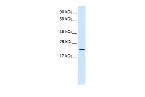
![Western blot using GeneTex's affinity purified anti-histone H2AvD pS137 antibody shows detection of a band at ~15 kDa corresponding to phosphorylated H2AvD (lane 2 arrow-head). Lanes contain either mock-irradiated (lane 1) or 4000-RAD gamma irradiated (lane 2) Drosophila melanogaster (3rd instar) larvae brain WC lysate separated on by SDS-PAGE and transferred to nitrocellulose. After blocking the membrane was probed with the primary antibody diluted to 1:500. Washes and reaction with secondary antibody followed incubation. Use HRP conjugated Gt-a-Rabbit IgG [H&L] MX and ECL for detection.](https://www.genetex.com/upload/website/prouct_img/normal/GTX48733/GTX48733_20160330_WB_w_23060823_592.webp)
- SizePrice
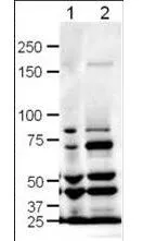
![Western blot using GeneTex's Affinity Purified anti-Mre11 antibody shows detection of a band ~80 kDa corresponding to mouse Mre11 (arrowhead). Lanes 1-4 contain 0.5 μg, 0.3 μg, 0.1 μg and 0.05 μg of purified mouse Mre11 protein, respectively. After 4-20% SDS-PAGE and transfer onto nitrocellulose, the membrane was blocked and then probed with the primary antibody diluted to 1:1,000 overnight at 4oC. The membrane was then washed and reacted with a 1:10,000 dilution of IRDye800 conjugated goat anti-Rabbit IgG [H&L] MX for 45 min at room temperature. IRDye800 fluorescence image was captured using the OdysseyR Infrared Imaging System developed by LI-COR. IRDye is a trademark of LI-COR, Inc. Other detection systems will yield similar results.](https://www.genetex.com/upload/website/prouct_img/normal/GTX48735/GTX48735_20160330_WB_w_23060823_965.webp)
- SizePrice
![Western blot using GeneTex's Affinity Purified anti-Rif1 antibody shows detection of a band ~265 kDa corresponding to mouse Rif1 (arrowhead). Specific reactivity with this band is blocked when the antibody is pre-incubated with the immunizing peptide (data not shown). Approximately 25 μg of MEF whole cell lysate was separated by SDS-PAGE and transferred onto nitrocellulose. After blocking the membrane was probed with the primary antibody diluted to 1.0 ug/ml for 2 h at room temperature followed by washes and reaction with a 1:10,000 dilution of IRDye800 conjugated goat anti-Rabbit IgG [H&L] MX for 45 min at room temperature. IRDye800 fluorescence image was captured using the OdysseyR Infrared Imaging System developed by LI-COR. IRDye is a trademark of LI-COR, Inc. Other detection systems will yield similar results.](https://www.genetex.com/upload/website/prouct_img/normal/GTX48737/GTX48737_20160330_WB_w_23060823_556.webp)
- SizePrice
- SizePrice
![Western blot analysis is shown using GeneTex's Affinity Purified anti-Swi6 antibody to detect endogenous protein present in S.pombe lysate (arrowhead). Comparison to a molecular weight marker (not shown) indicates a band of ~43 kDa corresponding to S.pombe Swi6 protein. ~35ug of lysate was loaded per lane onto a 4-20% gradient gel for SDS-PAGE followed by transfer to 0.45 mm nitrocellulose. The blot was incubated with a 1:1,700 dilution of the antibody at room temperature for 2 h followed by detection using IRDye?800 labeled Goat-a-Rabbit IgG [H&L] diluted 1:5,000 for 45 min. IRDye?800 fluorescence image was captured using the OdysseyR Infrared Imaging System developed by LI-COR. IRDye is a trademark of LI-COR, Inc. Other detection systems will yield similar results.](https://www.genetex.com/upload/website/prouct_img/normal/GTX48740/GTX48740_20160330_WB_w_23060823_421.webp)
- SizePrice
- SizePrice
- SizePrice
- SizePrice
- SizePrice
- SizePrice
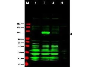
![Western blot using GeneTex's affinity purified anti-LDB1 antibody shows detection of LDB1 protein (arrowhead) in Jurkat whole cell lysate. Approximately 30 μg of lysate was loaded prior to separation and transfer to nitrocellulose. Primary antibody was used at a 1:1,800 dilution in 5% BLOTTO in PBS reacted overnight at 4oC. The membrane was washed and reacted with a 1:20,000 dilution of DyLight800 conjugated goat anti-Rabbit IgG [H&L] MX for 45 min at room temperature (800 nm channel, green). Molecular weight estimation was made by comparison to prestained MW markers in lane M (700 nm channel, red). Fluorescence image was captured using the OdysseyR Infrared Imaging System developed by LI-COR. IRDye is a trademark of LI-COR, Inc. Other detection systems will yield similar results.](https://www.genetex.com/upload/website/prouct_img/normal/GTX48750/GTX48750_20160330_WB_w_23060823_936.webp)
- SizePrice
- SizePrice
Didn't find what you were looking for?
Search through our product groups to find the right product
Back to overview