


Antibodies
We offer one of the most comprehensive portfolios of antibodies. This includes monoclonal and polyclonal primary, secondary, conjugated, phospho-specific, functional, (isotype) controls, tagged and antibody pairs. In addition, we offer custom antibody services from several manufacturers.
The antibodies are generated in various hosts and react to antigens of different species like human, mouse, rat, rabbit or zebrafish. The antibodies are validated for multiple applications, including immunohistochemistry, western blot, immunoprecipitation, ELISA and flow cytometry, to ensure reliable performance for your research needs.
If you need a specific antibody and can’t find it in our webshop, please contact our technical support.
Discover what our customers say about us by reading their reviews.
- SizePrice
- SizePrice
- SizePrice
- SizePrice
- SizePrice
- SizePrice
- SizePrice
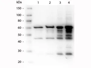
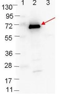
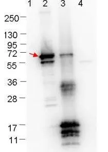
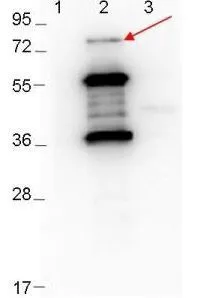
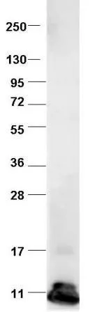
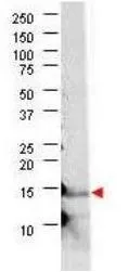
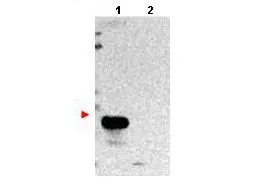
![Western blot using GeneTex's Protein-A Purified anti-bovine VEGF antibody shows detection of recombinant bovine VEGF-A at 17-19.2 kDa. Approximately 2 μg of recombinant protein was loaded per lane onto a 4-20% gradient gel followed by transfer to PVDF membrane. The membrane was blocked using 3% BSA diluted 1:10. The primary antibody was used at a 1:333 dilution and was incubated with the blot for 2h at room temperature. The membrane was washed and reacted with a 1:10,000 dilution of IRDye800 Conjugated Affinity Purified Goat-anti-Rabbit IgG [H&L] MX10. Molecular weight estimation was made by comparison to prestained MW markers. Other detection systems will yield similar results.](https://www.genetex.com/upload/website/prouct_img/normal/GTX48811/GTX48811_20160330_WB_w_23060823_126.webp)
- SizePrice
- SizePrice
- SizePrice
- SizePrice
- SizePrice
Didn't find what you were looking for?
Search through our product groups to find the right product
Back to overview